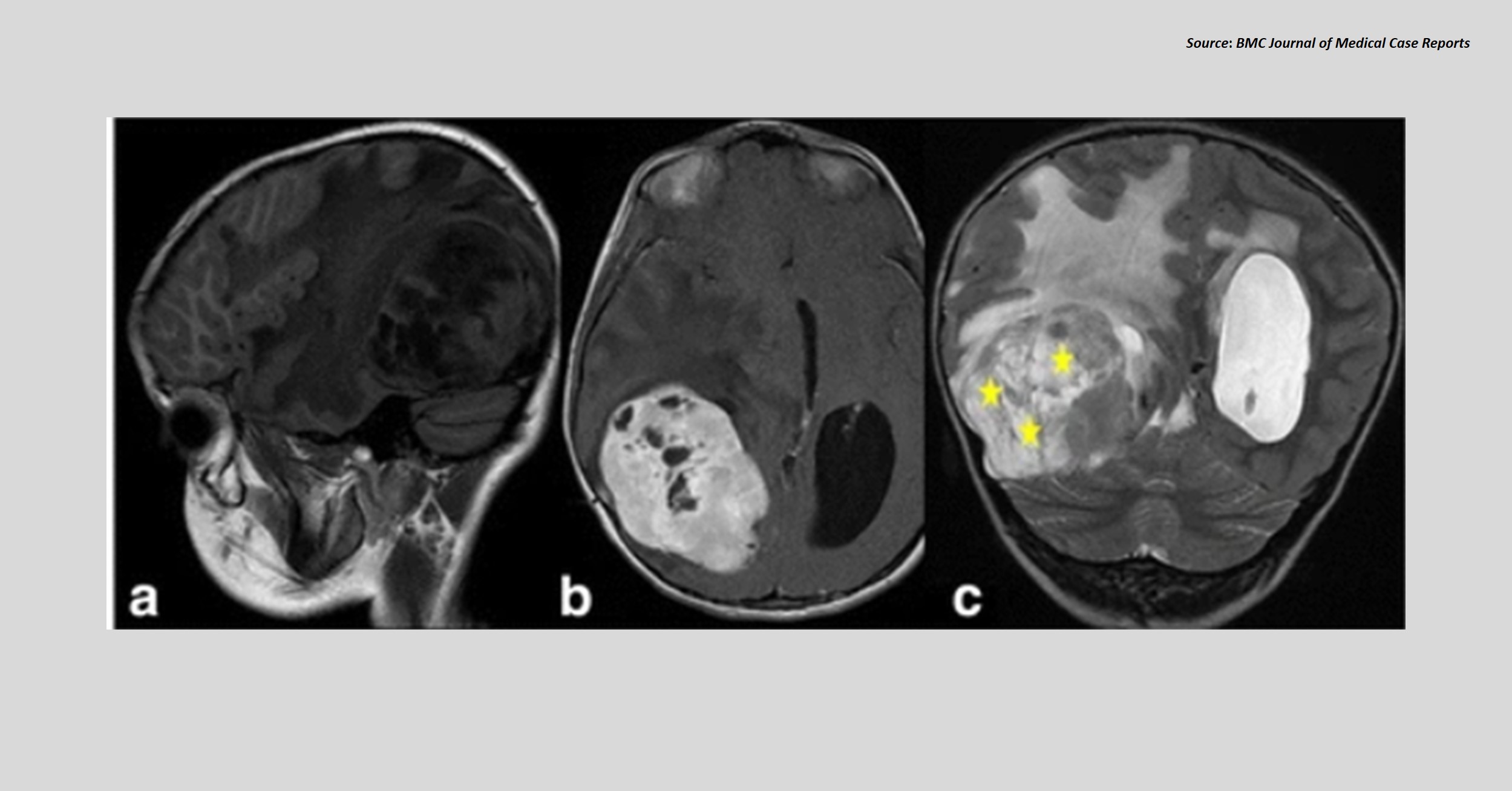
Case presentation An 8-year-old girl was presented to a hospital with a year-old history of headache, epileptic seizures, and decreased visual acuity. Diagnostic workup Cranial magnetic resonance imaging (MRI) revealed a well-demarcated voluminous mass at the right occipitotemporal region (Figures a, b, and c). The T2- and T1-weighted imaging revealed characteristic bubbly and hypointense heterogenous appearances, respectively. Resection and microscopic examination of the mass showed cells with indistinct cytoplasmic borders.
Immunohistochemical analysis revealed positive glial fibrillary acid protein (GFAP) staining (Figure d). Figure d. Immunohistochemical staining Source : BMC Journal of Medical Case Reports ##Disclaimer## The content provided on Docplexus is intended for educational purposes only. While we strive to ensure its accuracy, we make no warranties, explicit or…