
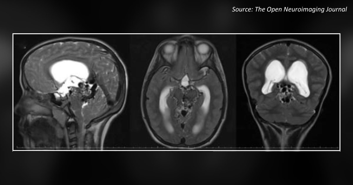
Case presentation An 11-year-old boy came to the outpatient department with complaints of progressive headache, nausea, and vomiting for the last month. The patient had been earlier investigated for right-sided facial palsy. Two years ago, he was diagnosed with an arteriovenous malformation (AVM) in the peri-mesencephalic region. Neurologic examinations: Bilateral papillary edema and right-sided central facial nerve palsy were observed. Radiological investigations: Head CT scan showed enlargement of lateral and third ventricles with transependymal cerebrospinal fluid oozing (Figure 1).
Cranial MRI revealed serpiginous signal void structures around the midbrain and enlargement of the Galen and internal cerebral veins (Cover Image). Figure 1. CT scan report Figure 2. Cerebral digital subtraction angiography In your opinion, what is the clinical diagnosis of the patient? Option A:…





