
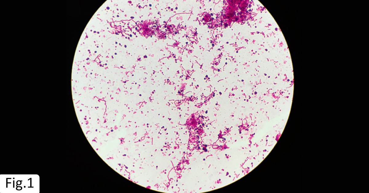
An 18-year-old man presented to the ED with a 1-week history of fever, sore throat, and malaise. The patient also reported a 3-day history of productive cough and pleuritic chest pain. He denied any history of intravenous drug use, known sick contacts, or recent travel. Clinical examination Febrile and ill-appearing While breathing ambient air, the oxygen saturation of patient was observed to be 88%. Diagnostic tests An anaerobic blood culture of the patient was initiated ( Fig. 1 ).
Radiograph analysis A chest radiograph of the patient revealed consolidation in the middle and upper portions of the left lung. A CT of the chest revealed multiple cavitary lesions ( Fig. 2A ), and a loculated left pleural effusion. A contrast-enhanced CT of the patient’s neck revealed an intraluminal filling defect in the right internal jugular vein ( Fig. 2B ). Diagnosis The patient was diagnosed with…





