
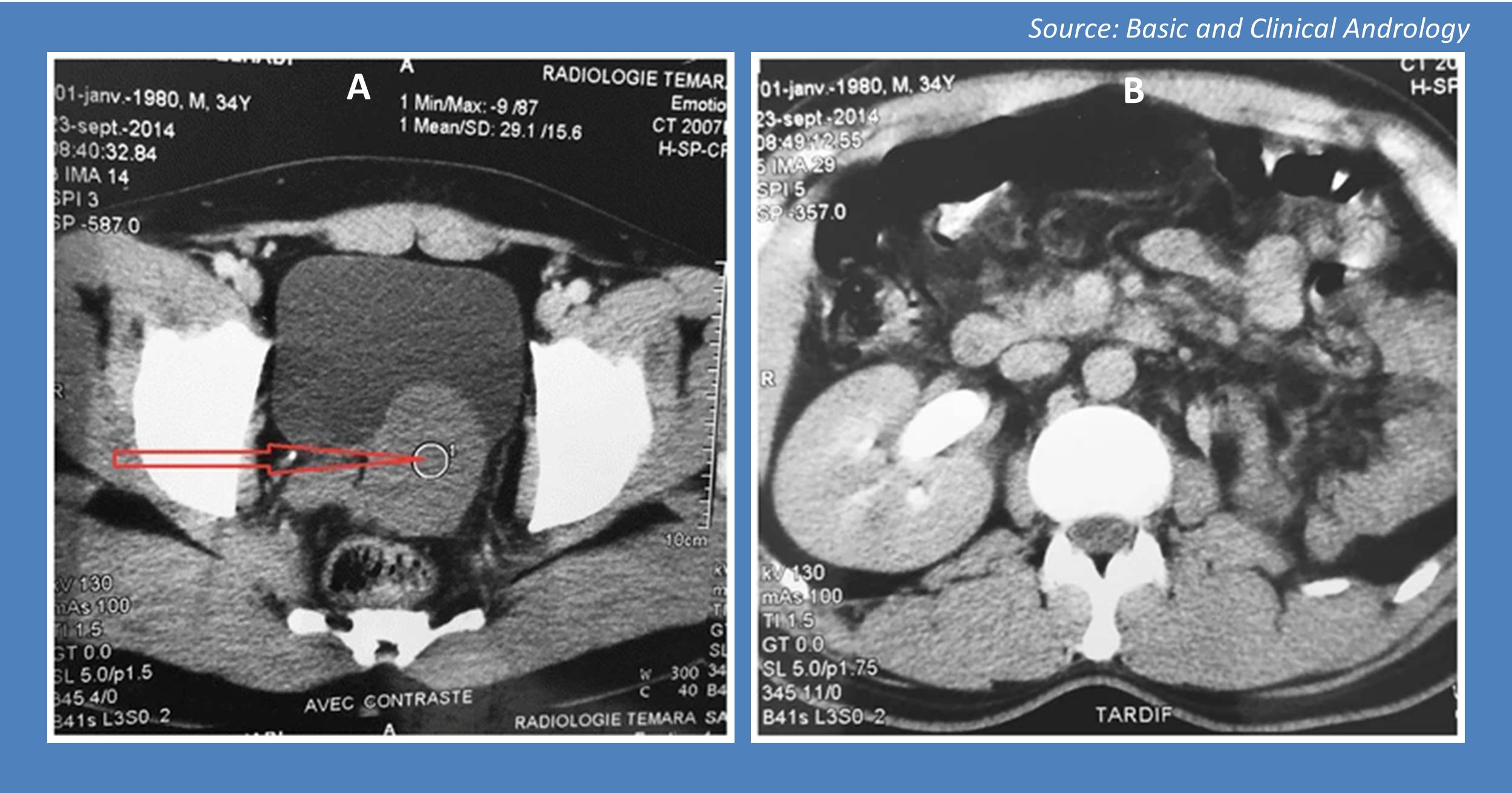
Case presentation A 35-year-old man hospitalized for intestinal subocclusion underwent an ultrasound of the abdomen that revealed a retrovesicular fluid-filled mass. Physical examination No evidence of palpable abdominal mass The biological assessment was unremarkable Radiological investigations 1. CT scan revealed – A homogeneous liquid mass measuring 80 x 60 mm with polylobed contours (Cover image A) Unilateral right-sided kidney (Cover image B) 2.
Pelvic MRI showed a single, thin-walled left-lateralized retrovesical, coarsely rounded measuring 56 × 54 × 44 mm (Figure 1) Source: Basic and Clinical Andrology This case is from the Editorial team of Docplexus and is meant for educative purposes only. For more such interesting clinical case challenges, click here!





