
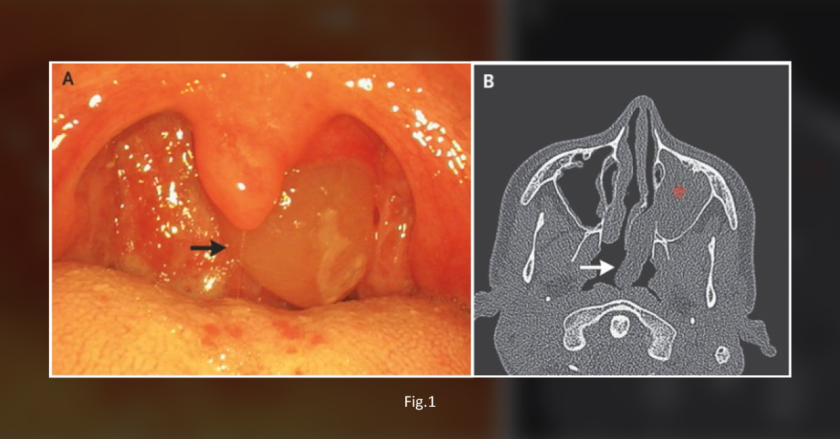
A 17-year-old boy presented to a clinic to get a pharyngeal mass evaluated. The patient did not report any other symptoms, such as rhinorrhea, nasal obstruction, epistaxis, or postnasal drip. Clinical examination An intraoral examination revealed a soft, translucent, round mass occupying the left pharyngeal area ( Figure 1A ). A rhinoscopy and CT scan of the sinuses revealed A mass originating from the left maxillary sinus ( Figure 1B ) The mass was observed to be extending through the accessory ostium that further protruded into the middle meatus and then exited posteriorly through the choana into the oropharynx.
Surgical procedure The patient was managed by endoscopic maxillary sinus surgery to remove the entire mass under general anesthesia. Histological Analysis Histological analysis of the mass was also carried out to confirm the diagnosis. Follow-up Post Surgery A follow-up visit…





