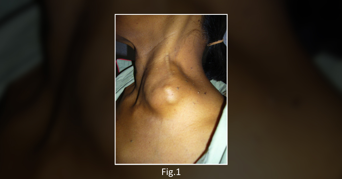
A 56-year-old woman presented to a clinic complaining of a painless mass in her left supraclavicular fossa and jaundice. The patient reported that the mass had progressively enlarged during the preceding eight weeks of this presentation. Physical examination The patient’s physical examination revealed: A hard lymph node which was about 6 cm by 6 cm in size located in the left supraclavicular fossa ( Figure 1 ) Hepatomegaly Radiograph analysis A plain radiograph of the patient’s chest revealed multiple nodular opacities in both lungs ( Figure 2A ) An abdominal CT scan revealed multiple lesions in the patient’s liver ( Figure 2B ) Endoscopic analysis An endoscopy of the patient’s upper gastrointestinal tract revealed a fungating mass present around the ampulla of Vater.
Biopsy analysis To initiate a biopsy analysis, a specimen was extracted from the mass and supraclavicular lymph. Based…