
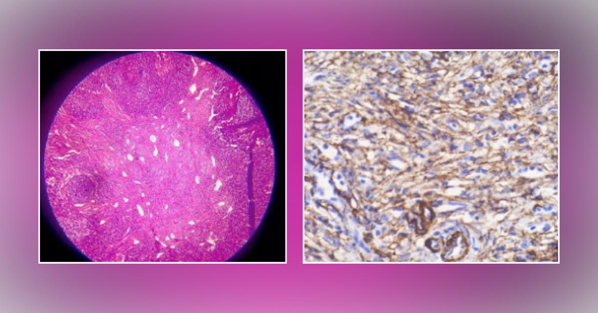
A 12-year-old girl was referred with the chief complaint of right inguinal mass since one year. The swelling was present with mild intermittent pain and slowly progressed to the current size. There was no history of chronic illness, trauma, weight loss or fever. Physical examination revealed a firm, mobile and mildly tender swelling, which was 4.5 × 4 cm in size. All laboratory investigations for tuberculosis were negative.
Microscopic examination of tissue biopsy exhibited A capsulated lymph node and parenchyma replaced by fibrous tissue containing numerous irregular blood vessels. Interspersed spindle cells and some lobules of benign adipocytes from hilum to cortex. Image is attached for reference. What is the most likely diagnosis? Option A: Lymphangiomatosis Option B: Angiomyomatous hamartoma Option C: Angiomyolipoma Option D: Vascular transformation of lymph node sinuses *This…


