
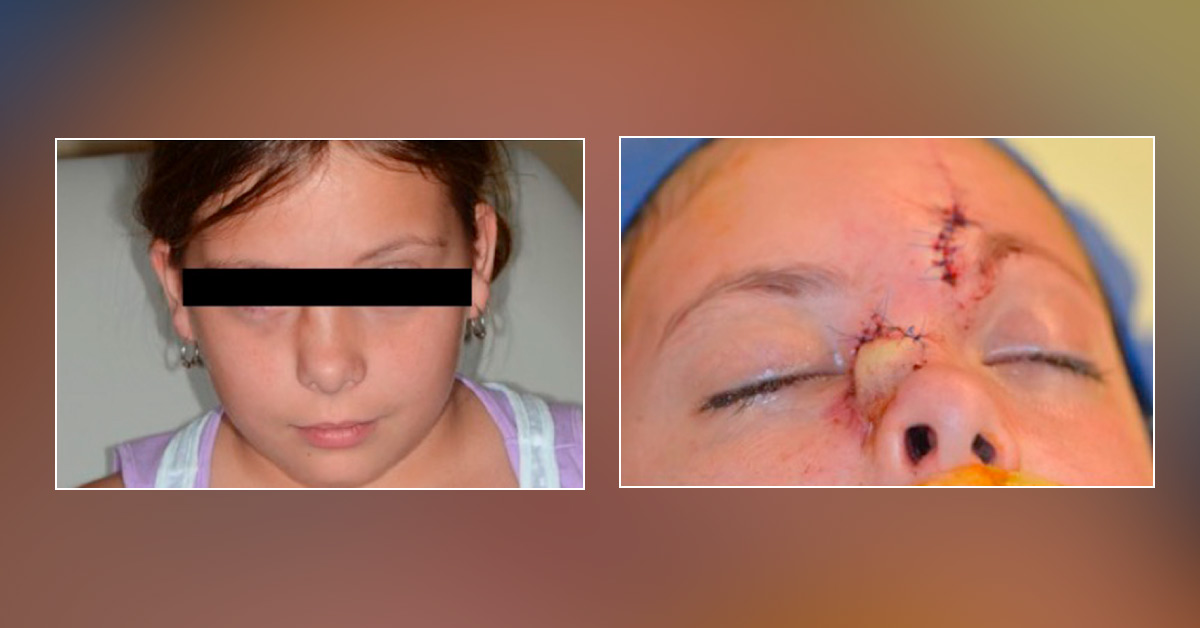
A ten-year-old female presented with an enlarging right nasal sidewall lesion resembling a persistent subcutaneous acne papule. The patient underwent endonasal excision of the mass; the diagnosis was unclear, and the specimen appeared to be a benign spindle cell lesion with a prominent vascular component. “Atypical spindle cell neoplasm” was diagnosed. Histological examination was remarkable for spindle cells with atypical tapering nuclei, an indistinct, pale cytoplasm, and local infiltration into skeletal muscle.
The morphological examination did classify as a known oncologic diagnosis, and immunochemical stains were negative for smooth muscle antigen (SMA), desmin, CD34, S100, and epithelial membrane antigen (EMA). The lesion recurred, and a second lesion developed adjacent to it; the masses were tender and did not exhibit overlying skin changes. Computed tomography and magnetic…





