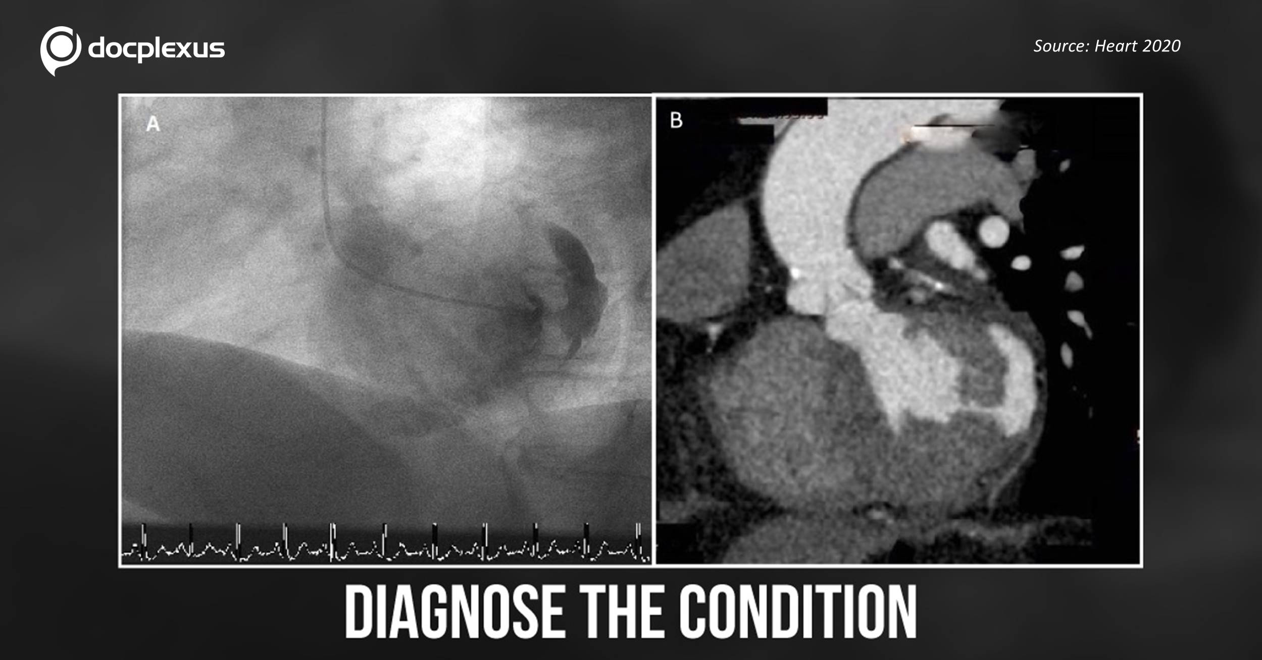
A 69-year old female patient presented with exertional dyspnea. She has a history of type 2 diabetes mellitus and hypertension for the past 15 years. Investigations 1. Transthoracic echocardiography Moderate to severe aortic stenosis Moderate aortic regurgitation Normal left ventricular (LV) systole Normal LV dimension 2. Left ventriculography (Figure A) Coronary arteries – normal A contrast filled pocket was observed on the lateral wall of the LV, synchronous with the LV.
3. Cardiac CT (Figure B) A radiodense pocket was visible continuous with the LV on its lateral aspect. ##Disclaimer## The content provided on Docplexus is intended for educational purposes only. While we strive to ensure its accuracy, we make no warranties, explicit or implied, about the suitability, reliability, or adequacy of the information, including data visualization elements and graphics. We do not endorse any…