
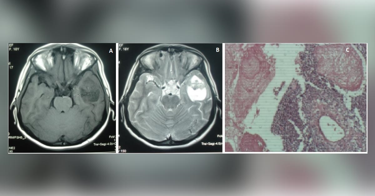
An 18-year-old woman with no past history of head trauma or surgery presented with a headache, dizziness, and blurred vision. Her physical and laboratory examinations were normal but neurological examinations revealed right homonymous hemianopia. MRI images showed large heterogeneous solid-cystic mass that had developed in the left temporal lobe. No significant edema or mass effect was observed.
Contrast-enhanced T1 weighted and T2 weighted images (A, B) and histopathologic examination (C) images have also been provided. What is the probable diagnosis? (Answer is posted in the comment section below!) *This case is from the Editorial team of Docplexus and is meant for educative purposes only.





