
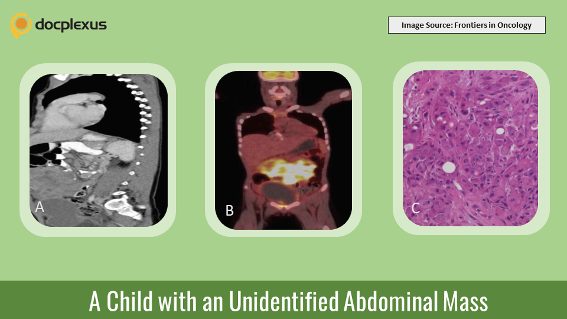
Case presentation An 8-year-10-month-old boy presents with abdominal pain, vomiting, constipation, and rectal pain for two weeks. A diffuse lower abdominal mass can be felt on palpation.
Investigations CT scan of abdomen – A large anterior multi-lobulated intraperitoneal mass, extending from above the umbilicus to the colovesical pouch (Cover image A) PET scan – High-grade fluorodeoxyglucose activity in the right lobe of the liver (Cover image B) Histopathological & peritoneal fluid assessment – Nests of pleomorphic epithelial cells with large nuclei and mitotic activity. The cells had rhabdoid features and cytoplasmic vacuoles (Cover image C) Immunohistochemical markers – No findings MRI of the bowel, CT scan of sinuses, USG of the scrotum, and endoscopies – Inconclusive Whole-exome sequencing – Missense mutations in the DNA-repair and chromatin-remodeling genes FANCM and SMARCD2 A…


