
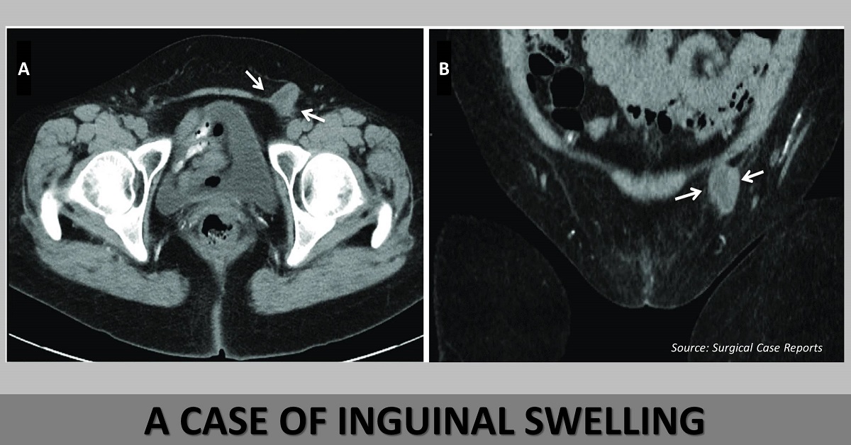
Case presentation A 56-year-old female presented with swelling of the left inguinal area that aggravated during activities. Physical examination Palpation revealed that the left groin mass did not return to the abdominal cavity. Radiological investigations Ultrasound (USG) of the lower groin revealed a cystic lesion , measuring 40 mm in diameter, which had unclear boundaries and appeared hypoechoic in the inguinal canal. Computed tomography (CT) scan showed that the cystic content was liquid , with irregular hypertrophy of the cyst wall (Cover image A).
The coronal section of the CT scan revealed that the cystic lesion did not communicate with the abdominal cavity (Cover image B). Surgical excision A laparoscopic surgical excision was performed under general anesthesia. Using an umbilical incision, a 12-mm trocar was inserted, along with two 5-mm working ports placed symmetrically. The…





