
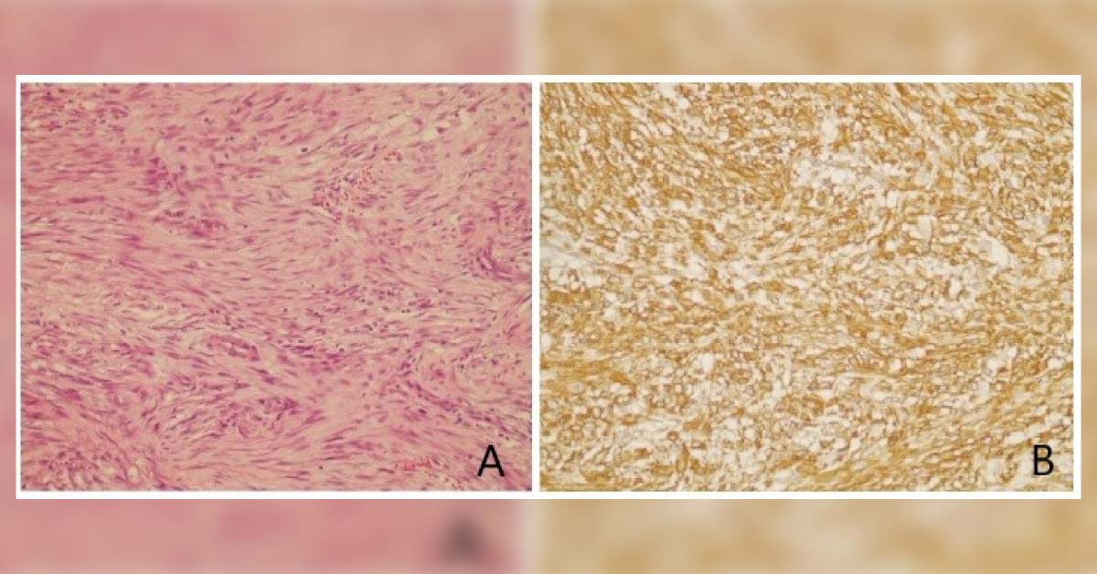
Case presentation A 48-year-old woman complained of a palpable mass and mild pain in her left breast and was admitted to the hospital. Family history She had no history of breast cancer in her family. Examination A small nodule was observed under the areola, which measured about 2 cm in diameter. Ultrasonography revealed a heterogeneous hypoechoic lesion in the breast parenchyma with infiltrative margins. It measured around 13 × 9 mm and extended to the subcutaneous tissue.
The lesion was also noted to have millimetric calcification foci. Pathology The mass was excised and found to have a measurement of about 2.5 × 2 × 2 cm. It had fragments of multiple irregular, white, soft tissue. Sectioning revealed a fibrous, white, and fatty lobular surface without any distinguishing figure. Microscopy images have been provided - hematoxylin and eosin (HE x40) staining (Image A) and smooth…


