
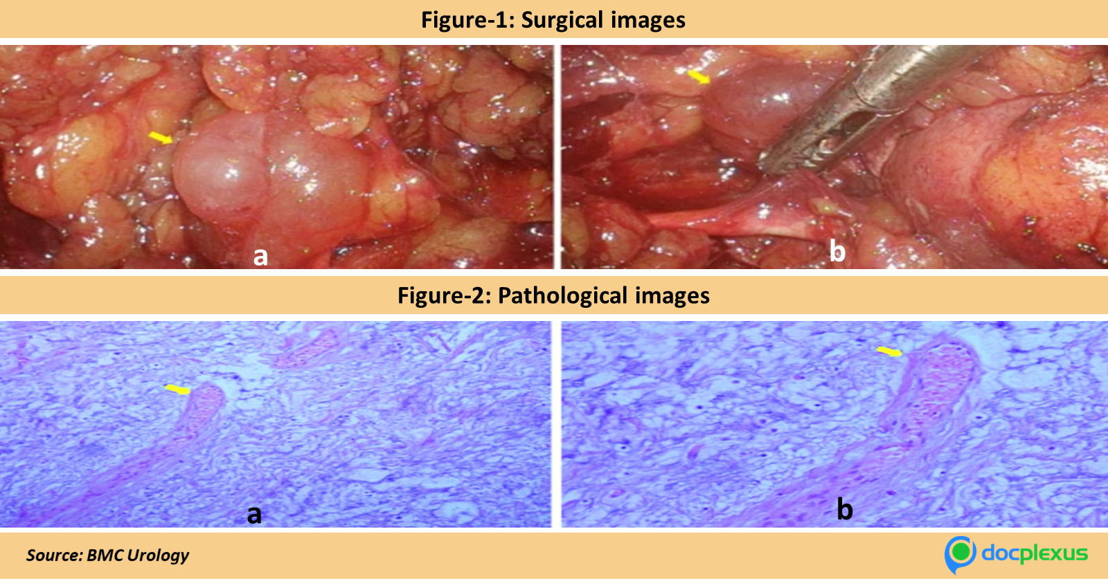
A 62-year-old male had a para-ureteral mass with a diameter of 15 mm, which was discovered accidentally during a routine check-up. No symptoms were identified. Abnormalities were absent when physically tested. Diagnostic findings: Surgical Images: A smooth, well-defined gelatinous cystic mass was observed with an inside tilt to the top-right ureter. The dimensions of the mass were at 2.3 x 2.0 x 1.0 cm. Refer to figure 1 for the surgical images.
Laparoscopic resection: Histopathological research found spindle-shaped cells containing variable calibre blood vessels in a loose myxoid stroma (figure 2). Computed tomography scanning: The left ureter had an abnormal mass 15mm in diameter. The mass was non-enhancing, with undefined boundaries on the ureter and hypodense on the muscle (figure 3). MRI: Displays hyperintensity on T2 weights (figure 4). What is the most likely diagnosis of this…


