
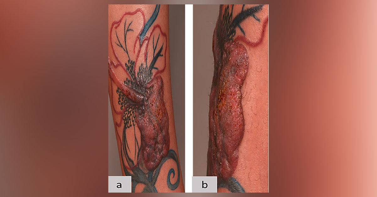
A 33-year-old woman, who had undergone a tattooing procedure nine months ago, presented with a massive infiltrated tumor that was livid red and itchy (Cover Image). Histopathology images have been provided (Figure 1)– (a) Hematoxylin and eosin (HE) staining, (b) CD3 staining, (c) CD4 staining, (e) CD30 staining, (f) CD20 staining, and (g) Ki-67 staining images. Figure 1. Histopathology images No clonal rearrangements of T-cell receptors could be detected in PCR. All other routine laboratory tests, including CBC, were normal.
What is your diagnosis? What treatment strategy do you recommend? *This image-based patient case is from the Docplexus Editorial Team for educative purposes only. The answer will be posted soon!



