
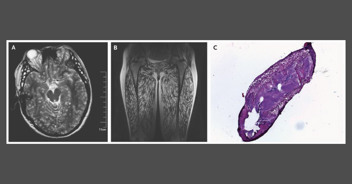
A 23-year-old woman presented with a one-month history of headache, vomiting, recurrent episodes of syncope, weight gain, and leg pain. A physical examination revealed several subcutaneous nodules on the patient’s face and trunk. Exophthalmos, fundal hemorrhage, neck rigidity, and a hypertrophic appearance of the thigh and calf muscles were also noted.
Magnetic resonance imaging (MRI) of the head (Image A) and Coronal MRI of the thighs (Image B) were performed. An oval translucent cyst was resected from the gastrocnemius muscle, and a histopathological examination of the cyst was carried out (Image C). Source: The New England Journal of Medicine





