
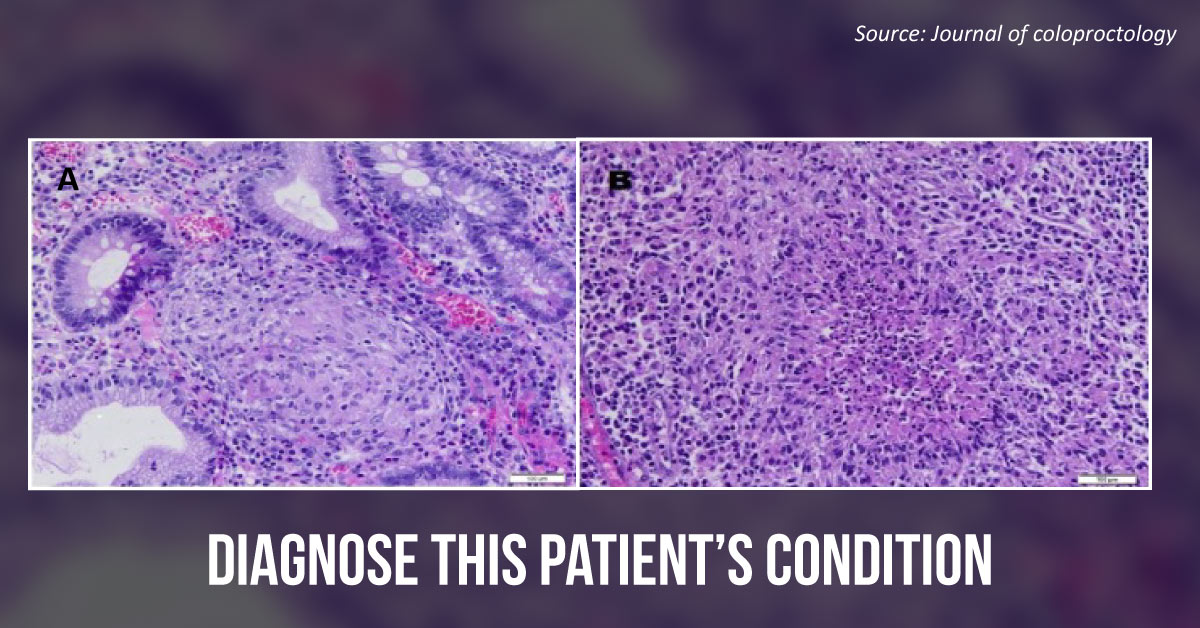
Case presentation A 26-year-old male animal care technician living in a rural area was admitted to the hospital with complaints of abdominal pain, diarrhea, and weight loss that had progressed over the past three years and worsened over the last few months. He presented with nausea, vomiting, and an acute obstructive abdomen with a palpable mass in the right iliac fossa. Radiological findings The CT scan revealed a big mass with a narrowed intestinal lumen and significant distention of the small bowel loops.
After retrograde intestinal cleansing , a colonoscopy was performed, which revealed severe inflammation of the cecum, preventing the colonoscope from progressing toward the terminal ileum. The patient underwent a right hemicolectomy followed by a side-to-side stapled ileocolonic anastomosis. Histopathological findings The surgical specimen analysis revealed non-necrotizing…



