
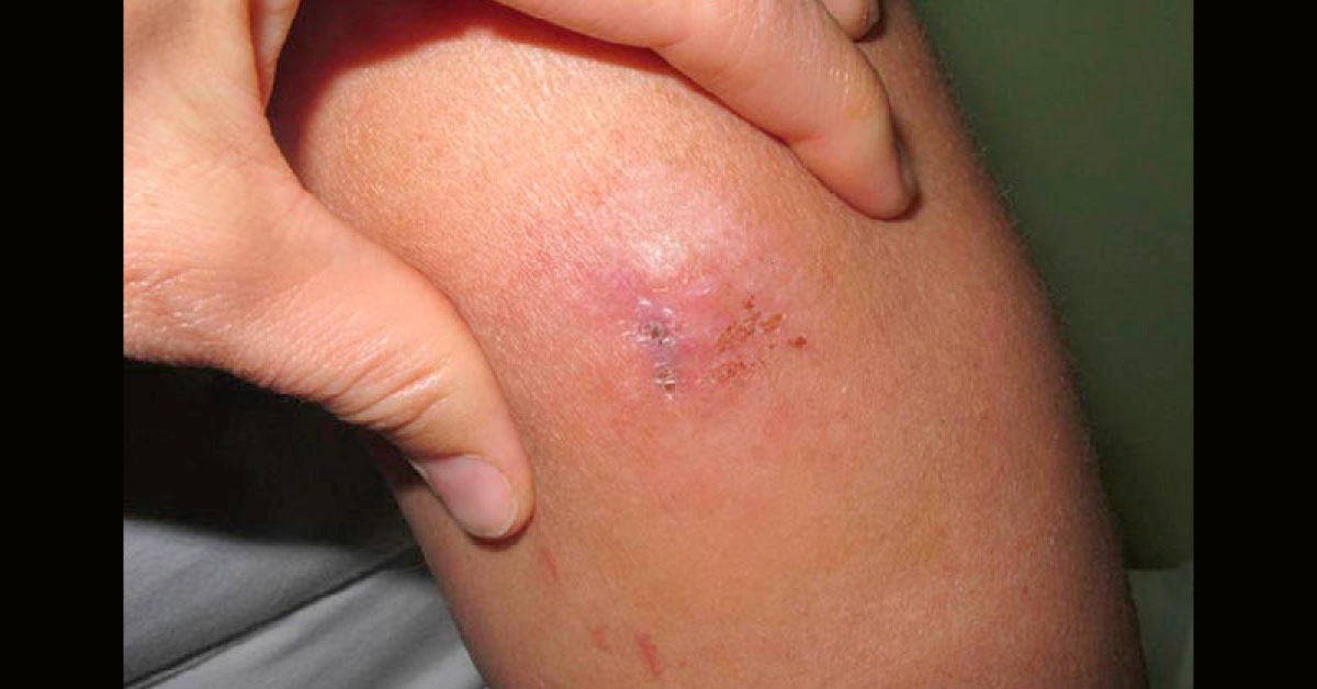
Patient History: A 31-year-old woman, with skin phototype IV and no relevant personal history, presented in our clinic with a painful progressively growing lesion that first appeared 1 year earlier. Physical Examination: Of note in the physical examination was the presence of a well-defined subcutaneous mass measuring 3×2.5cm (image attached). The lesion was of stony-hard consistency and infiltrated down to deep layers. Histological Findings: A biopsy was performed and sections were obtained for hematoxylin-eosin staining and immunohistochemical (IHC) staining.
Infiltration of the deep dermis and the subcutaneous cell tissue by small, uniform, cells were observed. These cells were arranged in a trabecular structure between a myxoid stroma (image attached). At higher magnification, a vesicular nucleus was observed in these cells with a high mitotic index. Large areas of necrosis were…