
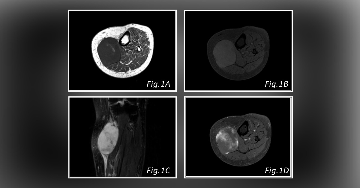
A 60-year-old female complained of a rapidly growing lump on her left calf. There was no pain, and the skin over the lump showed no change. Ultrasound finding : Vascularized mass on the left calf in the posterior muscular compartment MRI finding : For better characterization, MRI was performed. T1WI (Fig. 1A) showed high signal-intensity foci of fat within a principally low-signal-intensity mass. T1 with fat suppression imaging shows suppression of the signal in the foci that showed high signal on T1WI, which represented fat within the lesion (Fig. 1B) .
Sagittal T2FS (Fig. 1C) showed a hyperintense mass with some hypointense bands. There was exuberant soft tissue edema surrounding the lesion. The post-contrast study showed heterogeneous enhancement with a cotton-like appearance (Fig. 1D ) . Post-surgery complications: After surgery with complete resection and adjuvant chemotherapy, the…





