
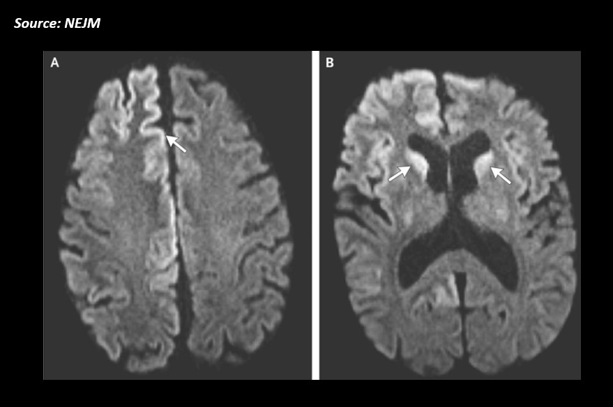
A 54-year-old male came to the emergency department with complaints of cognitive deterioration and functional decline for the last 20 days. He seemed disoriented, inattentive, and disinhibited. Neurologic examination: Horizontal gaze-evoked nystagmus, hyperreflexia on the left side, startle myoclonus, and ataxia.
EEG: Lateralized periodic discharges in the right hemisphere (with no evidence of seizures). Cerebrospinal fluid: Positive for 14-3-3 and tau proteins (known markers of prion disease) Radiological examination was conducted and the following observations were seen: Fig A: Hyperintensity of the cortical gyri of the frontal and occipital lobes, called cortical ribboning Fig B: Caudate heads on both the sides on diffusion-weighted imaging Source : NEJM ##Disclaimer## The content provided on Docplexus is intended for educational purposes only. While we strive to ensure its accuracy,…



