
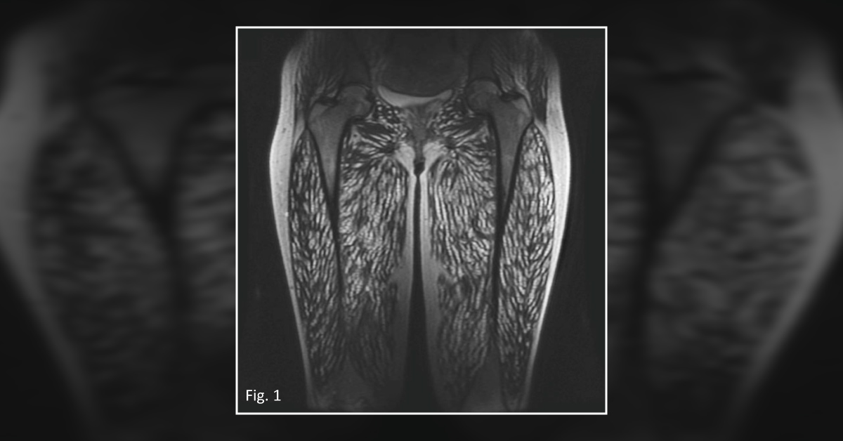
A 23-year-old female presented to a clinic with a 1-month history of vomiting, headache, weight gain, leg pain, and recurrent episodes of syncope. Physical examination The physical examination of the patient revealed: Multiple subcutaneous nodules distributed on the face and trunk Exophthalmos Fundal hemorrhage Neck rigidity A hypertrophic appearance of the thigh and calf muscles Radiographic analysis A coronal MRI of the patient’s thighs revealed numerous hyperintense lesions ( Fig. 1 ). An MRI of the patient’s head revealed diffuse hyperintense septated cystic lesions in the parenchymal, intraventricular, and retroocular regions ( Fig.
2A ). Other diagnostic test An ELISA assay was carried out on the serum and cerebrospinal fluid of the patient. Furthermore, an oval translucent cyst was also resected from the patient’s gastrocnemius muscle, and histopathological analysis was conducted…


