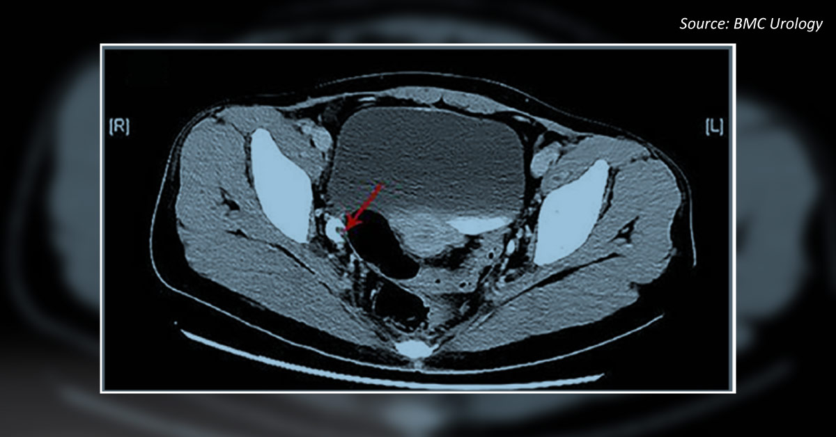
Case presentation A 37-year-old female visited the clinic, reporting intermittent hematuria with dysuria for the past ten days. However, there was no urinary urgency, waist discomfort, or fever. Physical examination The urethral orifice showed a soft, dark red strip-shaped protrusion with an undetermined root. Radiological examination A 36 x 17 mm mild hydronephrosis in the right kidney was observed in an ultrasound examination. A ureteroscope examination revealed that the base of the mass was in the lower and middle sections of the right ureter.
Computerized tomography urography (CTU) examination was performed (Cover image). Histological examination Immunohistochemistry was positive for GATA3 and negative for CK 20 cytoplasm. Based on the case presentation and CTU image, what is your most probable diagnosis? Inflammatory polyps Transitional cellular carcinoma Papillary urothelial…