
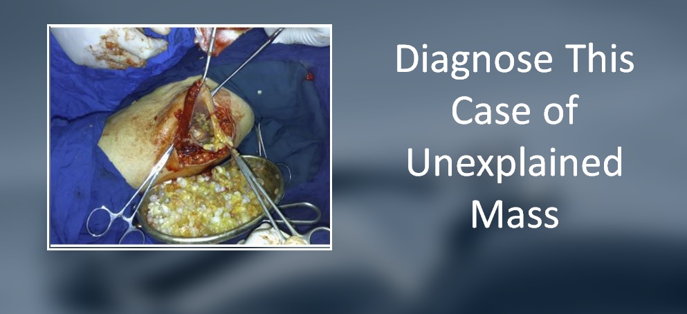
A 60-year-old woman presented to a surgeon for a painless mass in her right calf. She had the mass for the past two years, which later grew in size gradually. History The patient had a history of hypertension for the past ten years and was receiving hypertensive drugs. She had also undergone a laparoscopic cholecystectomy ten years ago and a hysterectomy for uterine fibroid five years ago. Investigation An examination revealed a soft mass in the posterior aspect of the right calf.
The mass measured 10 cm × 7 cm and was non-tender and mobile from side to side. An ultrasound of the mass showed a large cystic lesion containing several small daughter cysts (cover image). A chest X-ray excluded any pulmonary involvement. Additionally, an abdominal ultrasound excluded the involvement of intra-abdominal organs. This case is from the Editorial team of Docplexus and is meant for educational…





