
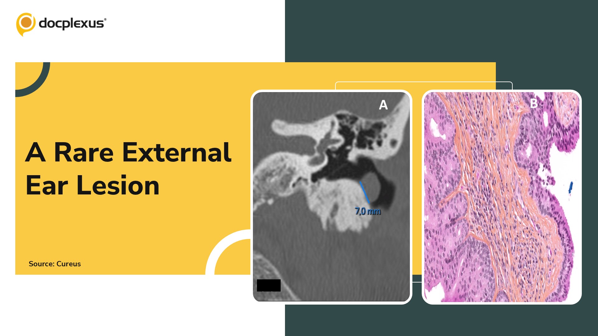
Case Presentation A 57-year-old man presented with a two-year history of intermittent left-sided ear fullness associated with recurrent cerumen impaction. He denied otalgia, otorrhea, tinnitus, or hearing loss.
Otoscopic Examination Soft, smooth, flesh-colored pedunculated mass arising from the posterior wall of the left external auditory canal (EAC), partially obstructing the canal Audiometry: Normal bilateral hearing thresholds Imaging Temporal bone CT (non-contrast): • 6 mm pseudonodular cutaneomucosal lesion at the entrance of the left EAC with minimal underlying osteolysis (~2 mm) of the posterior canal wall (Cover Image A) • Tympanic membrane intact, no middle ear involvement Surgical Findings Approach: Transcanal excision under local anesthesia Intraoperative findings: • Serous fluid aspirated from the lesion • Lesion excised completely down to its base • Small erosion of…



