
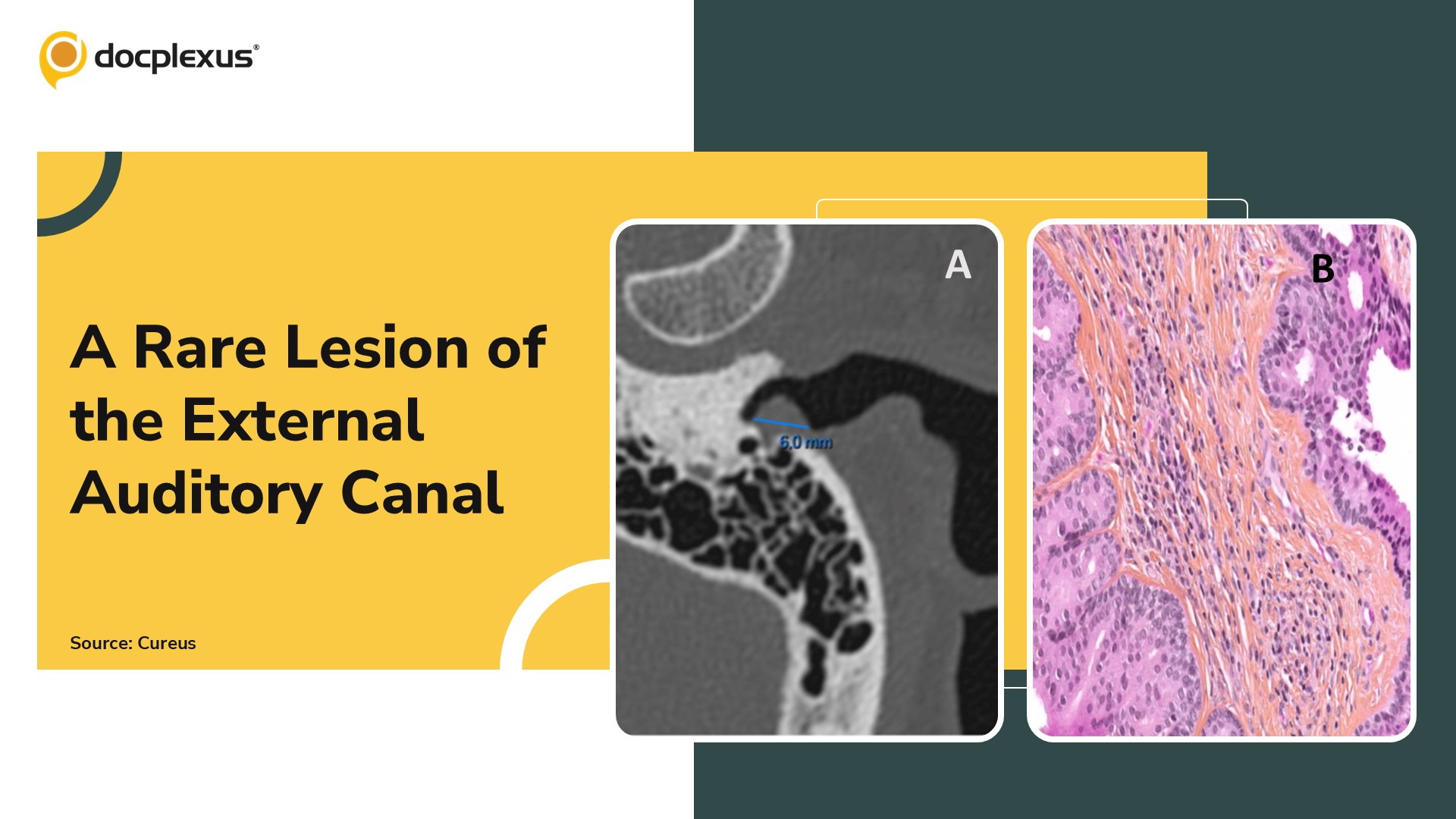
Case Presentation A 57-year-old man with no significant medical or surgical history presented with a two-year history of intermittent left-sided ear fullness and recurrent cerumen impaction. He denied otalgia, otorrhea, tinnitus, or hearing loss.
Initial Examination Otoscopic evaluation revealed a soft, smooth, flesh-colored pedunculated mass arising from the posterior wall of the left external auditory canal (EAC), partially obstructing the canal Audiometry demonstrated normal bilateral hearing thresholds Based on these findings, a benign obstructive lesion such as a ceruminous adenoma or papilloma was provisionally suspected Imaging Findings Temporal bone computed tomography (CT): Demonstrated a 6 mm pseudonodular cutaneomucosal thickening at the entrance of the left EAC Associated with minimal underlying osteolysis (~2 mm) of the posterior canal wall (Cover Image A) Tympanic…



