
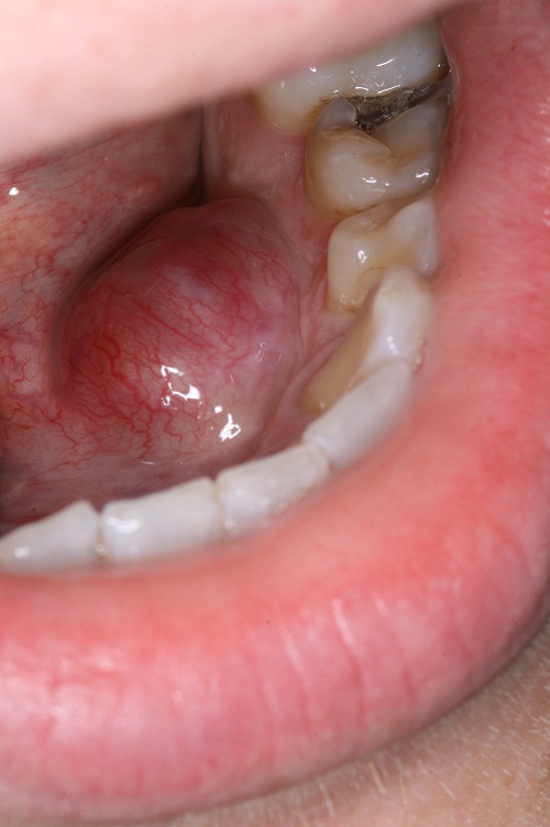
A 27-year-old patient presented with a five-year history of an enlarging mass in the left side of the floor of mouth. It was exophytic and smooth surfaced with prominent surface vasculature (Image 1 attached). On palpation, the nodule was firm . The color was pink to slightly red. There was no ulceration or purulent drainage. The swelling was about 1.5 cm in diameter.
Under IV sedation, the lesion was completely excised. The lesion was firm to palpation. The area healed with no complications. An excisional biopsy was ordered (Images attached). Image 2: Screening power (x40) H & E stained section shows a large and encapsulated neoplasm demonstrating two patterns of histology (Antoni A & Antoni B) plus obvious cystic degeneration, hypocellularity and bleeding in the center of the neoplasm. Image 3: Low-power (x100) H & E stained section demonstrating bundles of spindle-shaped cells…