
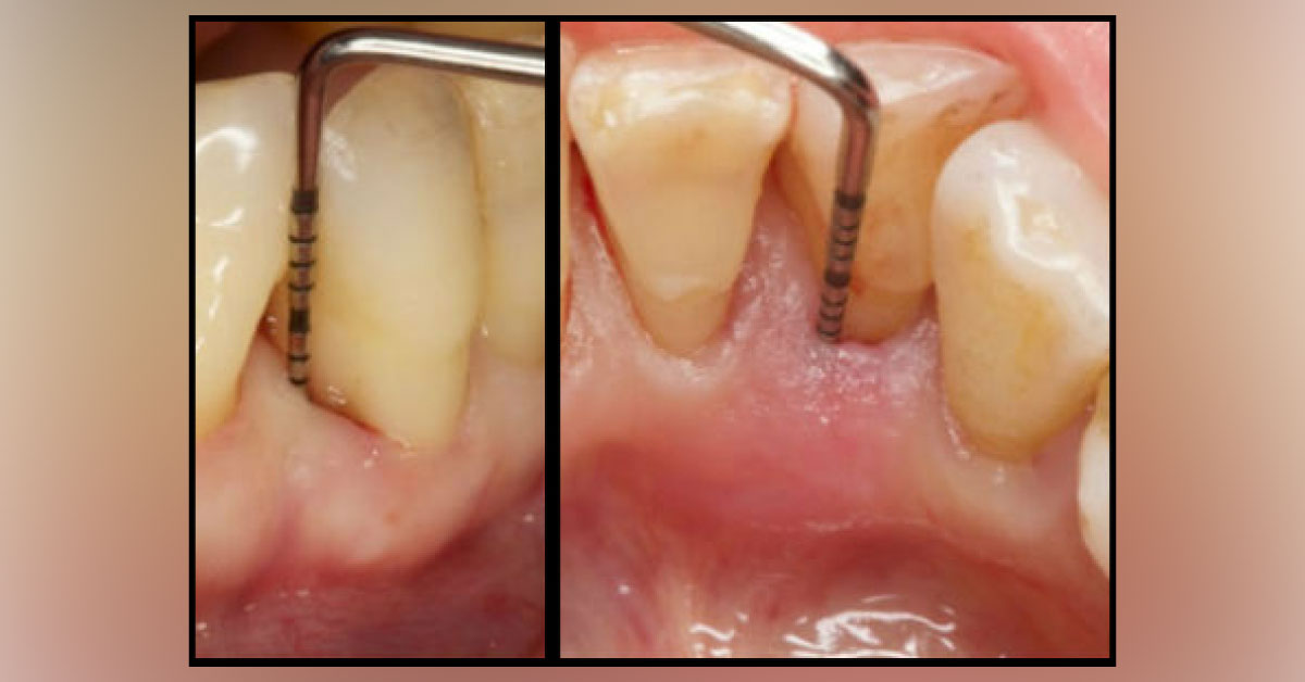
A 71-year-old female was referred to the Department of Periodontology for a periodontal exam. The patient denied any current and past medical history, allergies and medications. Upon clinical and radiographic exam (Image 1 a and b), the patient was diagnosed with generalized slight to moderate, with localized severe, chronic periodontitis on tooth #26. Clinical findings included marginal erythema, BoP, 7 mm PPD on the lingual of #26. Gingival recessions were observed on teeth #25, 26 and 27.
Tooth #26 was rotated with its distal margin facing the labial. Grade 2 mobility and fremitus were noticed on #26. Image 1 : Clinical aspect of tooth #26, buccal (a) and lingual (b) view of the probing depths Image 2 : Radiographic image of tooth #26 in a lateral (a) and frontal (b). *This case is from Docplexus editorial team for educative purpose only Source: Trends in Clinical Periodontology and…