docplexus
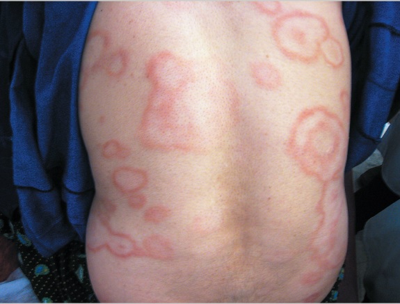
A 30-year-old woman presented with a 4-month history of a rash on her back. The physical examination revealed multiple large, annular, hypopigmented, atrophic macules with well-defined, erythematous, raised borders. The lesions were hairless, hypohidrotic, and anesthetic and had developed at a slow and progressive pace. There was no peripheral-nerve enlargement. Histopathological analysis of a skin-biopsy specimen revealed well-developed epithelioid granulomas, lymphocytes, and Langerhans' cells surrounding neurovascular structures within the papillary dermis.
Dermal nerves were swollen and destroyed. No acid-fast bacilli were detected on modified Ziehl–Neelsen staining. What could be the possible diagnosis? What treatment course would you suggest? *This case is from the Docplexus Editorial Team and is meant for educative purpose only.