
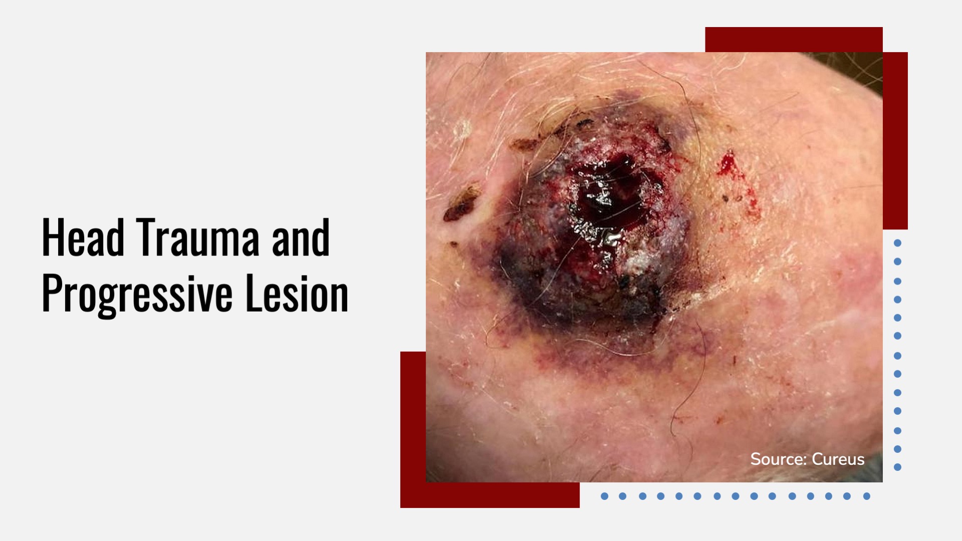
Case presentation A 90-year-old Caucasian male veteran presented to a geriatric clinic with head trauma (Cover image). He had a 3 cm hematoma and a bruise on the head. Medical history Alzheimer’s dementia Squamous cell carcinoma of the scalp and lung (stage I), which was treated with resection Chronic obstructive pulmonary disease Disease progression One month after the initial presentation, the patient presented with bleeding at the scalp trauma site. Cellulitis was suspected, and the patient was treated with cephalexin without healing the lesion.
Another month later, bleeding and ulceration developed with a lesion (2.5 cm diameter) consisting of granulation tissue and pearly borders. CT scan Swollen tissue (3.7 cm diameter) was found in the superior posterior portion of the left frontal area. The maximum thickness of the tissue was about 13 mm. Intact calvarium without foreign bodies…


