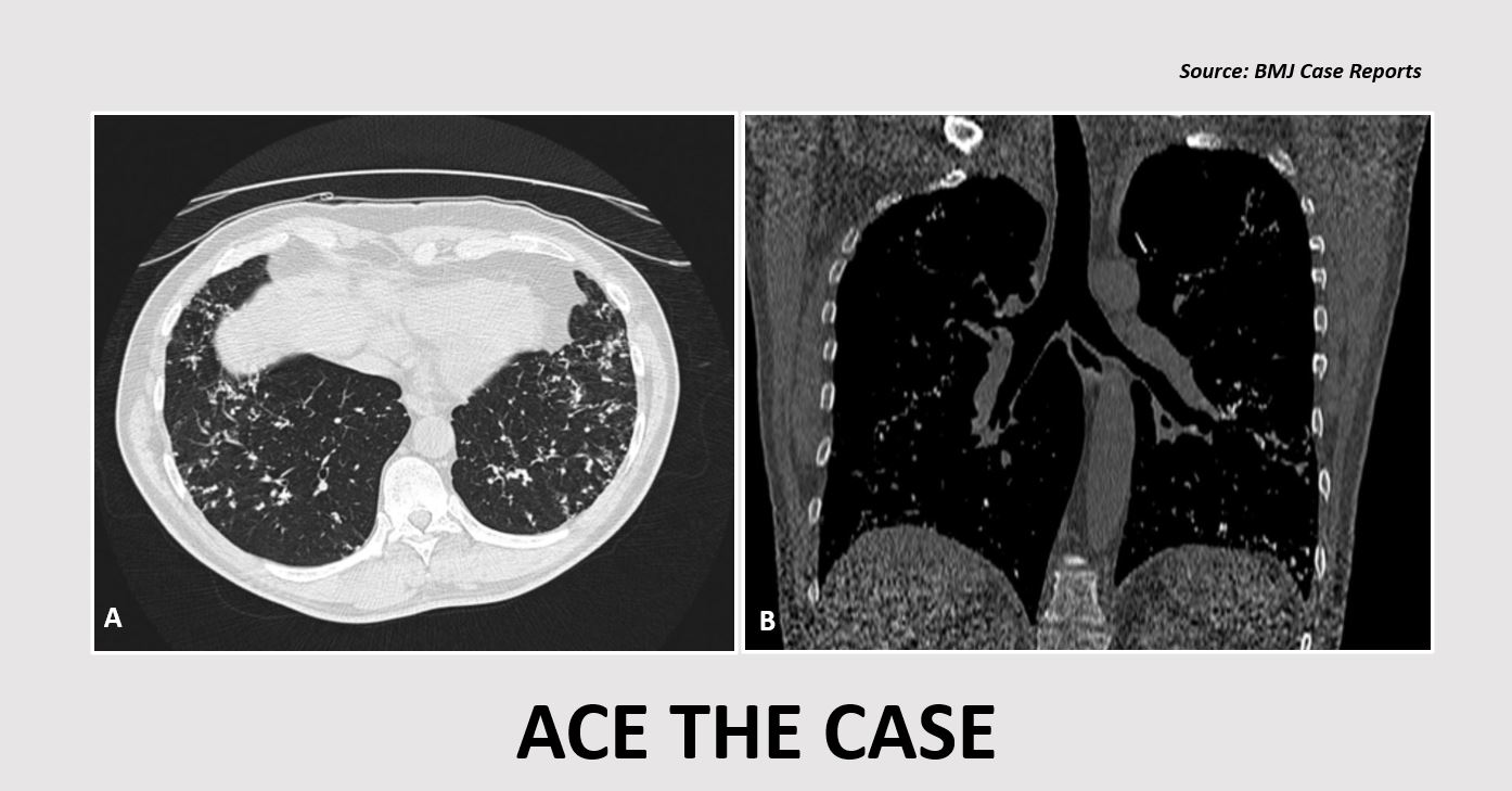
Solve this exciting clinical case specific to your field of interest via Docplexus’ Clinical Case Challenge. Join your fellow doctors to discuss and answer today’s challenge. Case presentation A 47-year-old man was referred to a hospital due to an abnormal chest X-ray (CXR). The patient was asymptomatic with no significant family history.
Radiological findings Chest X-ray revealed: The current CXR findings were similar to the CXR taken ten years ago and showed bilateral interstitial markings suggestive of pulmonary fibrosis. High-resolution CT scan (HRCT) revealed: Multiple bilateral intra-alveolar depositions of sand-like calcifications, predominantly in the lung bases (Cover images A and B). Ground-glass opacities in the lingula No enlarged mediastinal lymph nodes Spirometry No abnormalities were detected. Forced expiratory volume in 1 second - 102% Forced vital capacity - 118%…