
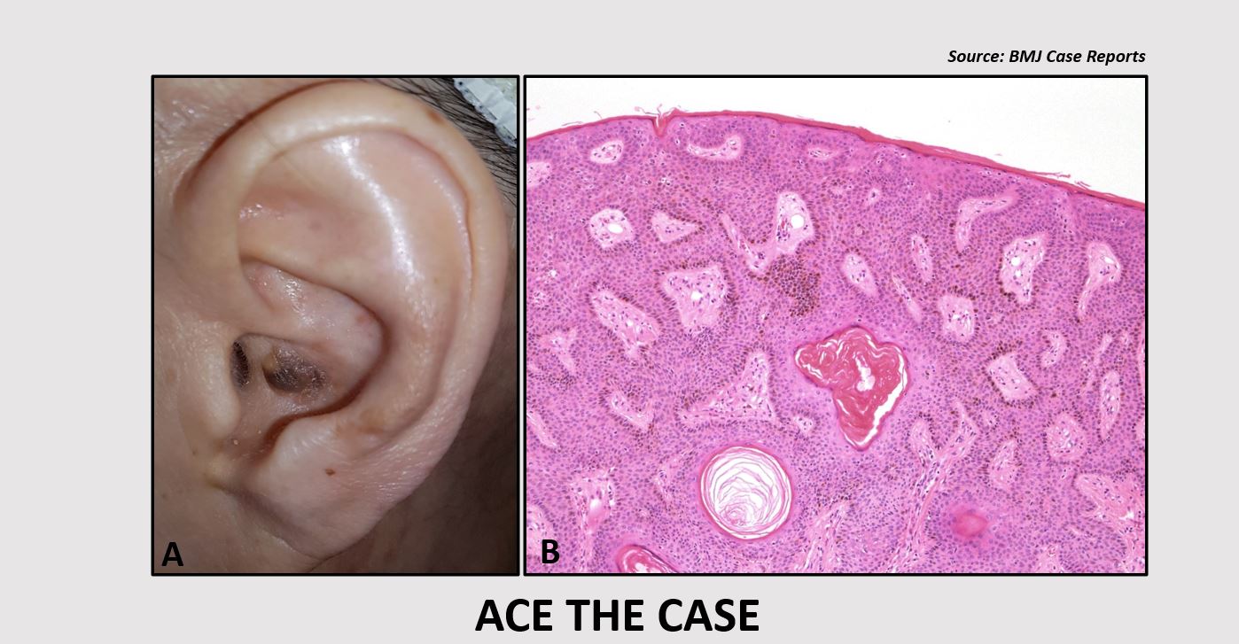
Case presentation A 69-year-old woman reported to the otorhinolaryngology department with a mass in the left auricle. The mass had been present for several years, and no pain or itching was associated with it. The patient reported no history of trauma or overexposure to ultraviolet light. Physical examination A brownish verrucous papillomatous lesion (1 x 0.5 cm) was observed in the cavum concha of the left auricle (Cover image A). The doctors suspected malignancy. The lesion was excised completely and sent for a histopathological evaluation.
Histopathological findings (Cover image B) Basal cell proliferation in the epidermis Papillomatosis, hyperkeratosis, and acanthosis Based on the physical examination and histopathological findings, what is your probable diagnosis for this patient? A. Keratoacanthoma B. Seborrheic keratosis C. Verruca vulgaris D. Actinic keratosis *This image…