
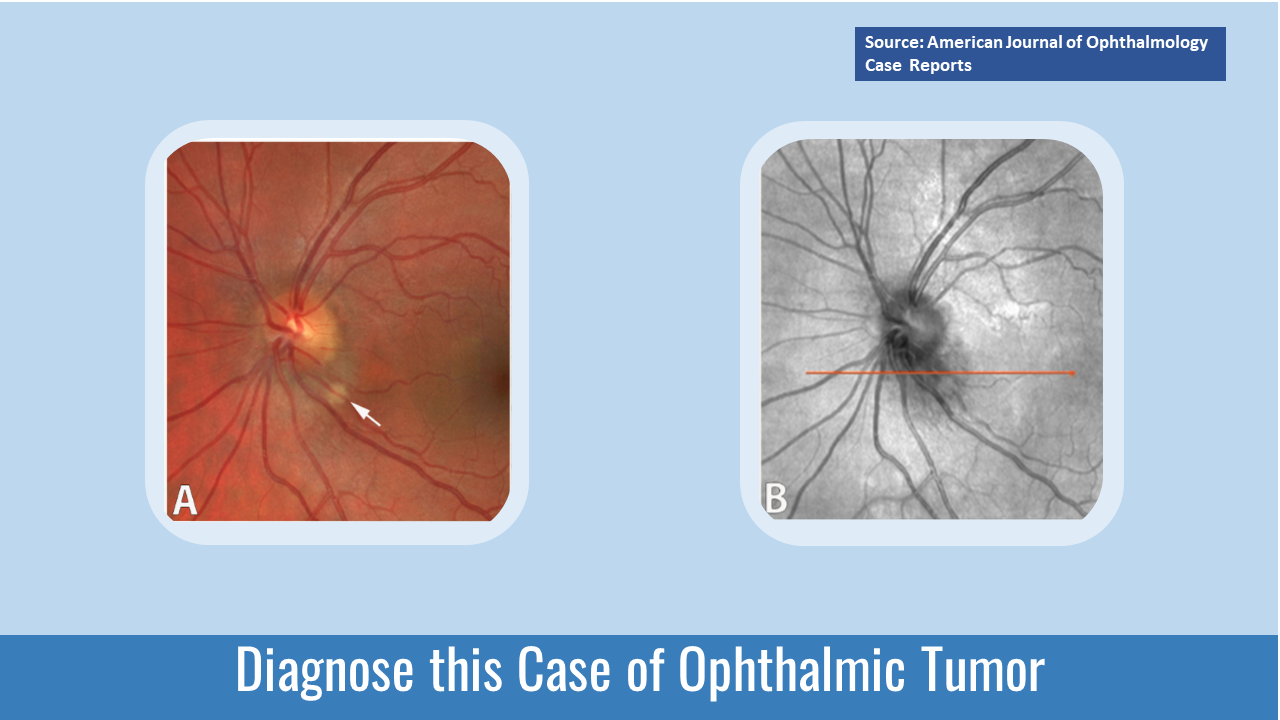
A 30-year-old female with a family history of Von-Hippel-Lindau disease presented to the Ophthalmology Department of a hospital for a routine checkup. Examination Vision in the left eye — 20/20-1 Vision in the right eye — 20/400 Color fundus photography — Peripapillary lesion- hypopigmented spot inferotemporal to the optic disc (Cover Image A) Optical computed tomography (OCT) of the optic disc —Hyporeflective area inferotemporal to the optic nerve head (Cover image B) . Lesion—2.2 mm (Figure 1) Cross-section OCT — Disrupted retinal fibers & ellipsoid zone No disruption of the retinal pigment epithelium near the tumor Swept-source optical coherence tomography angiography (SS-OCTA) scan (3x3 mm) of the lesion was carried out to visualize the lesion.
Images were segmented manually between the retinal pigment epithelium and the inner plexiform layer. Two vascular foci were identified in…



