
Case presentation: A 19-year-old man presented with shortness of breath and complained about losing weight continuously for several months. Radiological investigation: A chest radiograph showed a large anterior mediastinal mass from the manubrium to the diaphragm. The mass was solid and heterogeneous, displaced the great vessels, and narrowed the main bronchi. Echocardiography revealed a large pericardial effusion with cardiac tamponade.
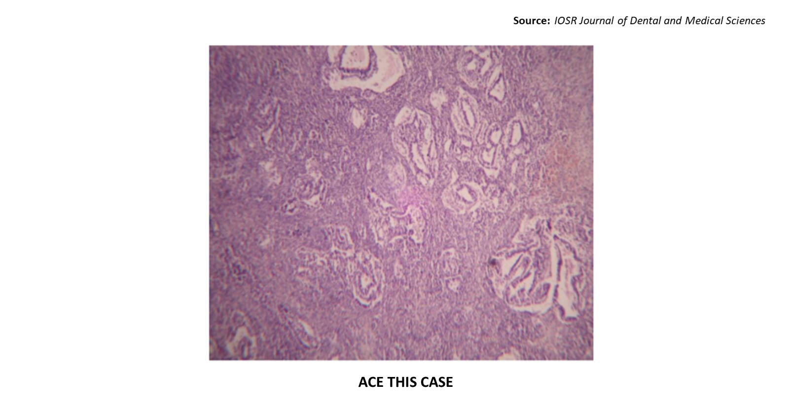
Laboratory investigations: An open biopsy of the mediastinal mass showed a solid neoplasm with a glandular architecture. It consisted of highly atypical cells with a moderate amount of eosinophilic cytoplasm and large, round nuclei with prominent nucleoli (Cover image). Source : IOSR Journal of Dental and Medical Sciences ##Disclaimer## The content provided on Docplexus is intended for educational purposes only. While we strive to ensure its…


