
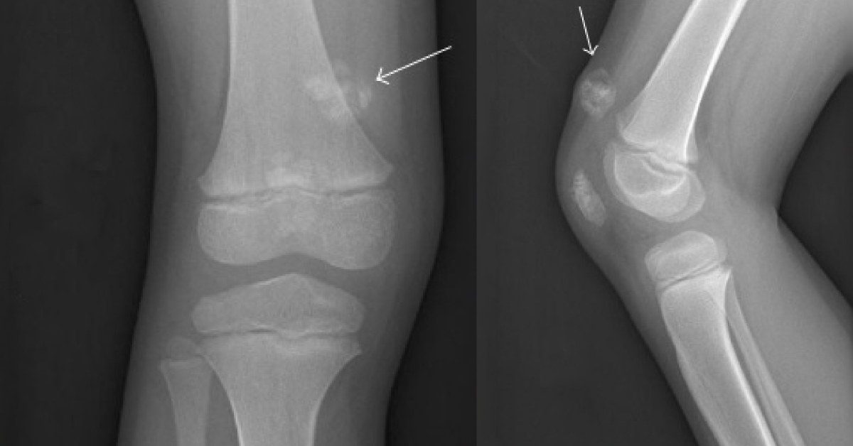
A 5-year-old female was presented with a chief complaint of a mass in the anterior distal right thigh. The mass was noted to be slow growing but unknown when it first appeared. There were no complaints of pain, no history of trauma in the region, and no other lesions identified. On physical examination, the mass was firm and well adhered to the dermis but freely mobile over the underlying fascia.
There were no skin changes or discoloration and the knee exam was otherwise normal. Radiological investigations Plain radiographs showed an oval, calcified mass within the soft tissue of the suprapatellar region (images attached). Magnetic resonance imagining revealed a mass confined to the subcutaneous soft tissues along the anteromedial aspect of the thigh superficial to the vastus medialis causing extrinsic mass effect without deep invasion This mass was predominantly low signal intensity…





