
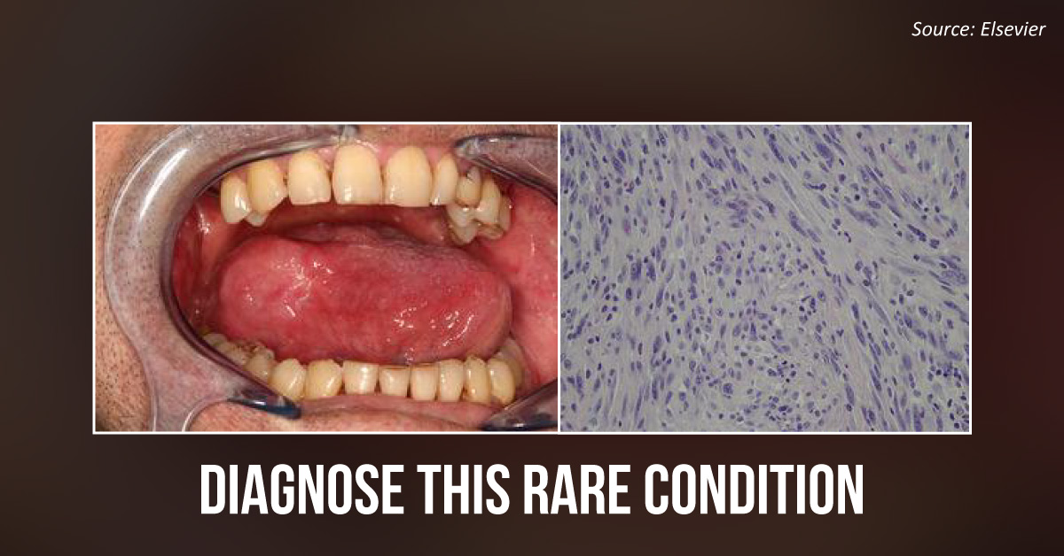
Case presentation A 40-year-old male patient was referred to the maxillofacial department after he complained of a rapidly increasing lesion on the right side of his tongue. Medical history Two years prior to this presentation, the patient underwent radiotherapy and chemotherapy because of a Cavum Carcinoma. Clinical examination Oral examinations revealed a well-circumscribed, firm, submucosal mass on the right side of the tongue. Cervical lymphadenopathy was observed during the examination. However, the mass was not palpable.
CT scan The scan demonstrated a submucosal mass on the right side of the tongue. After a previous incisional biopsy turned out to be negative for carcinoma, a repeat biopsy was conducted. The results of the histopathological examination of the biopsy samples are shown in the cover image. The microscopic examination showed interlacing fascicles of spindle-shaped…





