
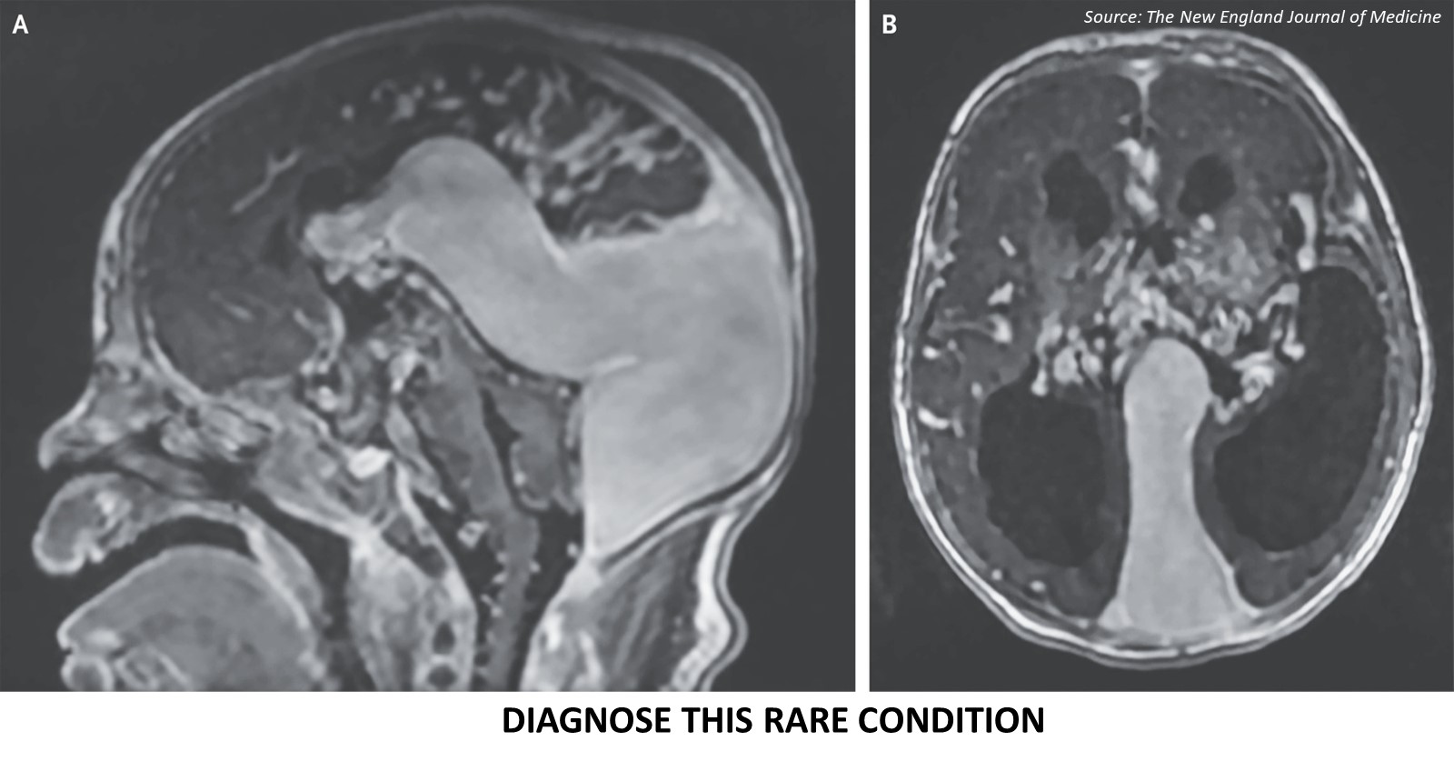
Case presentation: A neonate born at 37 weeks of gestation experienced respiratory distress three hours after birth. Physical examination: Subcostal retractions, coarse breath sounds in both lungs, and a systolic murmur were evident. Radiological investigation: A chest radiograph showed signs of cardiomegaly and bilateral pleural effusions.
An echocardiogram revealed a patent foramen ovale, a dilated right atrium, a dilated and hypertrophied right ventricle, tricuspid regurgitation, and right-to-left shunting at the patent ductus arteriosus. Cranial ultrasonography and gadolinium-enhanced T1-weighted MRI scan showed a large choroidal type of malformation, hydrocephalus, and atrophy of the brain stem and cerebellum in the sagittal (Cover image A) and axial (Cover image B) views. Source : The New England Journal of Medicine ##Disclaimer## The content provided on Docplexus is intended for…


