
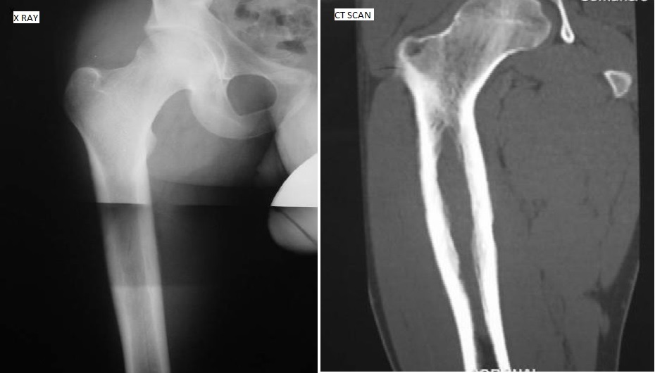
Sixteen-year-old boy with pain in the right hip and thigh for several months. He has no history of fevers, night sweats, weight loss. There was no previous history of infections. Laboratory studies including CBC, ESR and CRP were normal. X-ray: X-rays demonstrated a permeative lesion of the right proximal femur with slight sclerosis. The lesion was barely perceptible on the Xray. CT scan: The CT scan demonstrated a permeative lesion through the proximal ½ of the femur.
The cortex was mildly thickened and expanded. There was no soft tissue component. MRI: The T1 weighted MRI demonstrated a permeative lesion involving the upper ½ of the femur. The T2 weighted image demonstrates significant edema. What could be the possible diagnosis on the basis of radio imaging? *This case is by Docplexus Editorial Team for educative purpose only.





