
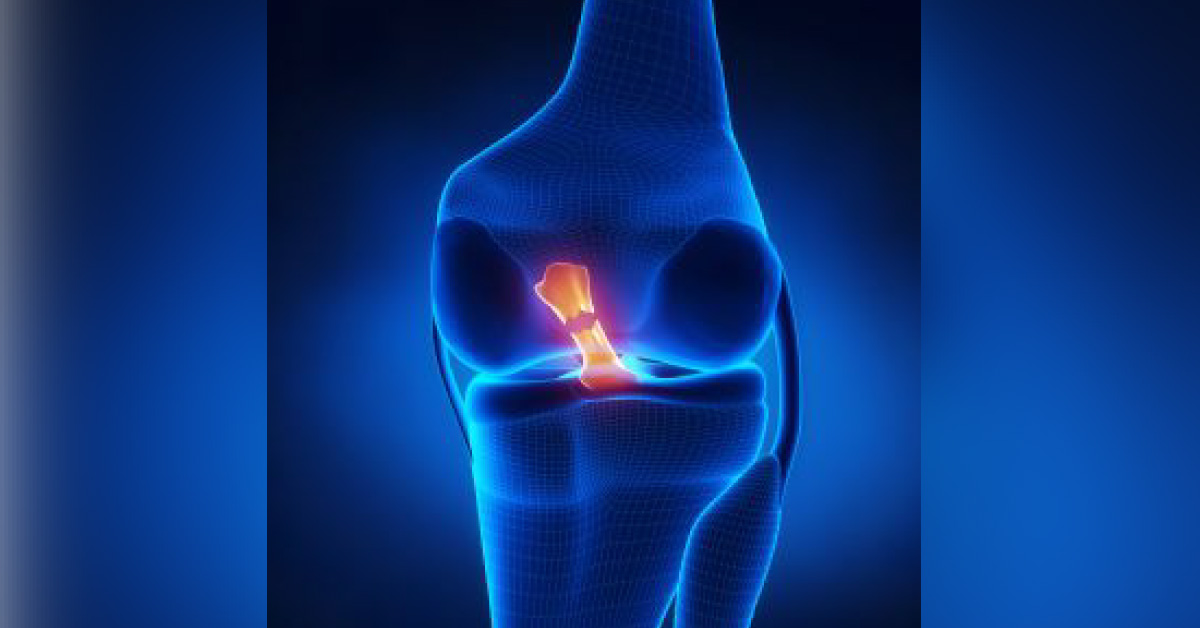
The anterior cruciate ligament (ACL) is an important structure that maintains the normal biomechanics of the knee and commonly injured knee ligament. However, the oblique course of the ACL within the intercondylar fossa limits the visualization and assessment of the pathology of the ligament. This article outlines anatomy and different modalities of radiological investigation of ACL. The anterior cruciate ligament (ACL) runs in an oblique course from the tibia to the lateral femoral condyle.
It is an intra-articular extra synovial ligament composed of fibres running from the anterior intercondylar region of the proximal tibia to the medial aspect of the lateral femoral condyle within the intercondylar groove. Anatomy The fibres of the ACL are arranged into two bundles known as the anteromedial and posterolateral bundle according to their tibial insertion. The anteromedial bundle inserts…





