
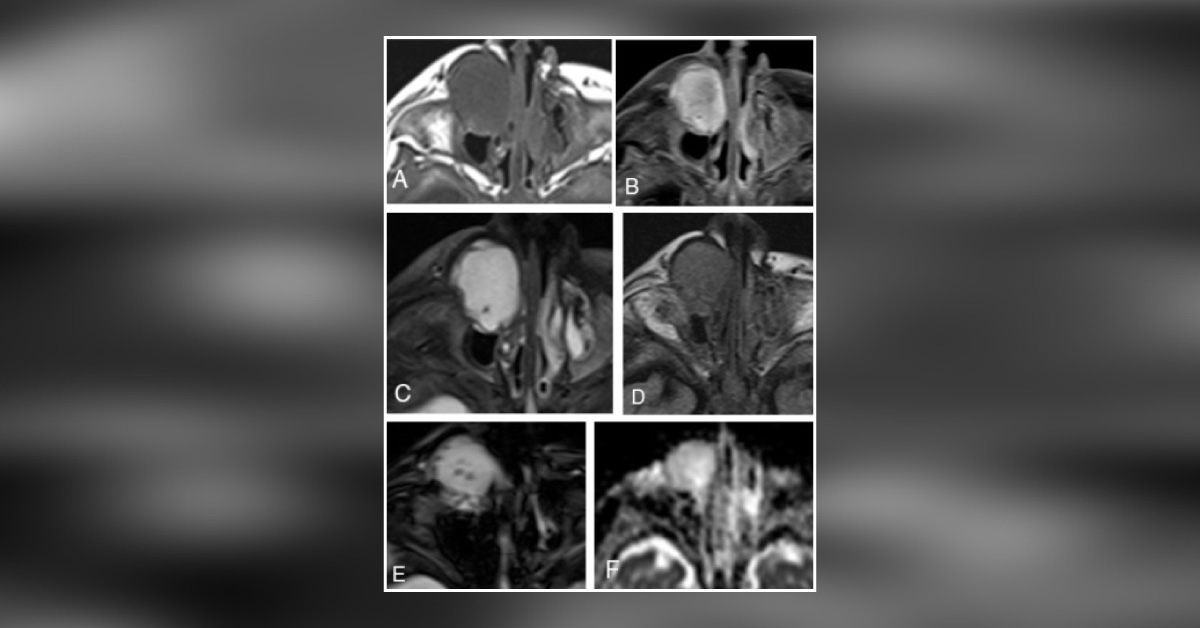
Figure A. T1W unenhanced axial image: Homogeneous T1 hypointensity in a well-circumscribed, low-attenuation mass over the right nasolacrimal gland. B. Avid homogeneous enhancement on the post-contrast, fat-saturated T1 view. C. Homogeneous T2 prolongation throughout the lesion except for a punctate focus of low T2 signal posteriorly.
D. FLAIR-weighted images-intermediate signal (lack of fluid suppression), solid or proteinaceous material. E. GRE axial images - small oval susceptibility artifacts of degraded blood products or mineralization. F. ADC map in the axial plane - high signal within the lesion, facilitated diffusion.





