docplexus
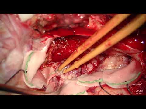
41 year old man presented with history of progressive right external ophthalmoplegia over 6 months. Examination revealed complete right external ophthalmoplegia. Imaging revealed a large mass lesion involving the middle cranial fossa and extending through the tectorial incisor into the posterior fossa. The origin of the tumour appears to be tectorial incisor and then extending to the middle and posterior fossa.
He underwent endovascular embolisation of the tumor using PVA particles followed by sub temporal approach and total resection of the middle fossa portion of the tumor. Lumbar drain was placed during surgery to achieve brain relaxation.