
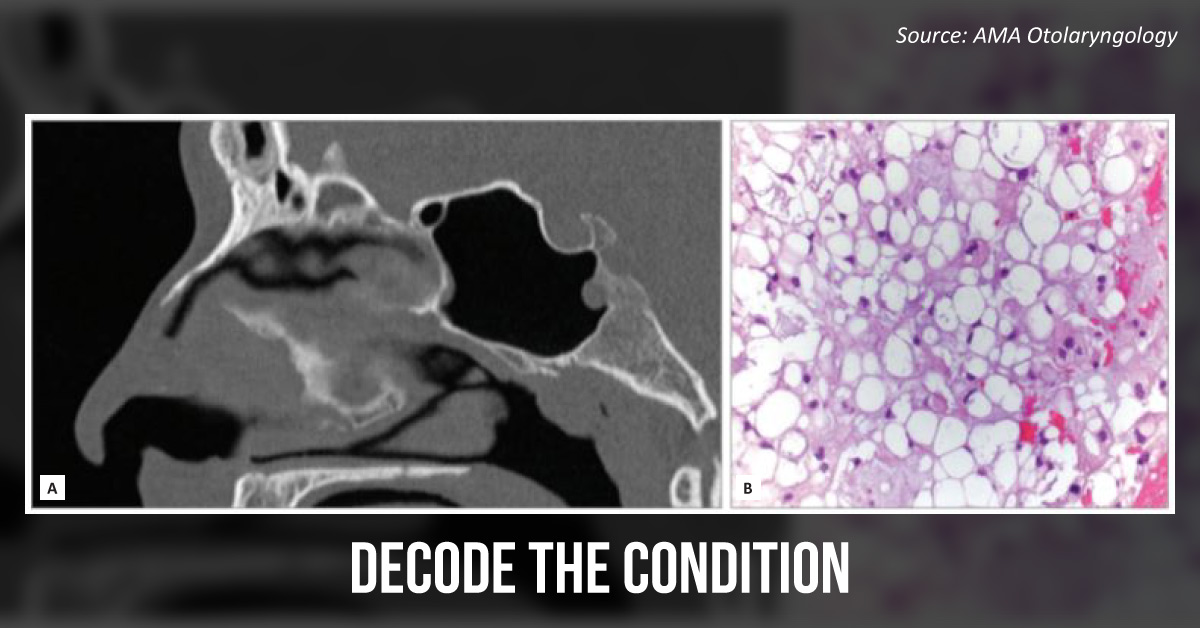
Case presentation A young man presented to the ED with facial trauma post an assault. Physical examination marked tenderness over the nasal bones. CT scan (without contrast) of the bone showed a fracture of the nasal bone and a 7 mm tissue density within the posterior aspect of the left sphenoid sinus (Image A).
An MRI with contrast revealed a transosseous, multilobulated mass of size 2.1 × 1.4 × 1.3 cm with heterogenous enhancement extending into the sphenoid sinus. Pathological examination of the tissue biopsy specimen showed sheets of neoplastic, vacuolated, soap-bubble-like cell structure with a myxoid background (Image B).





