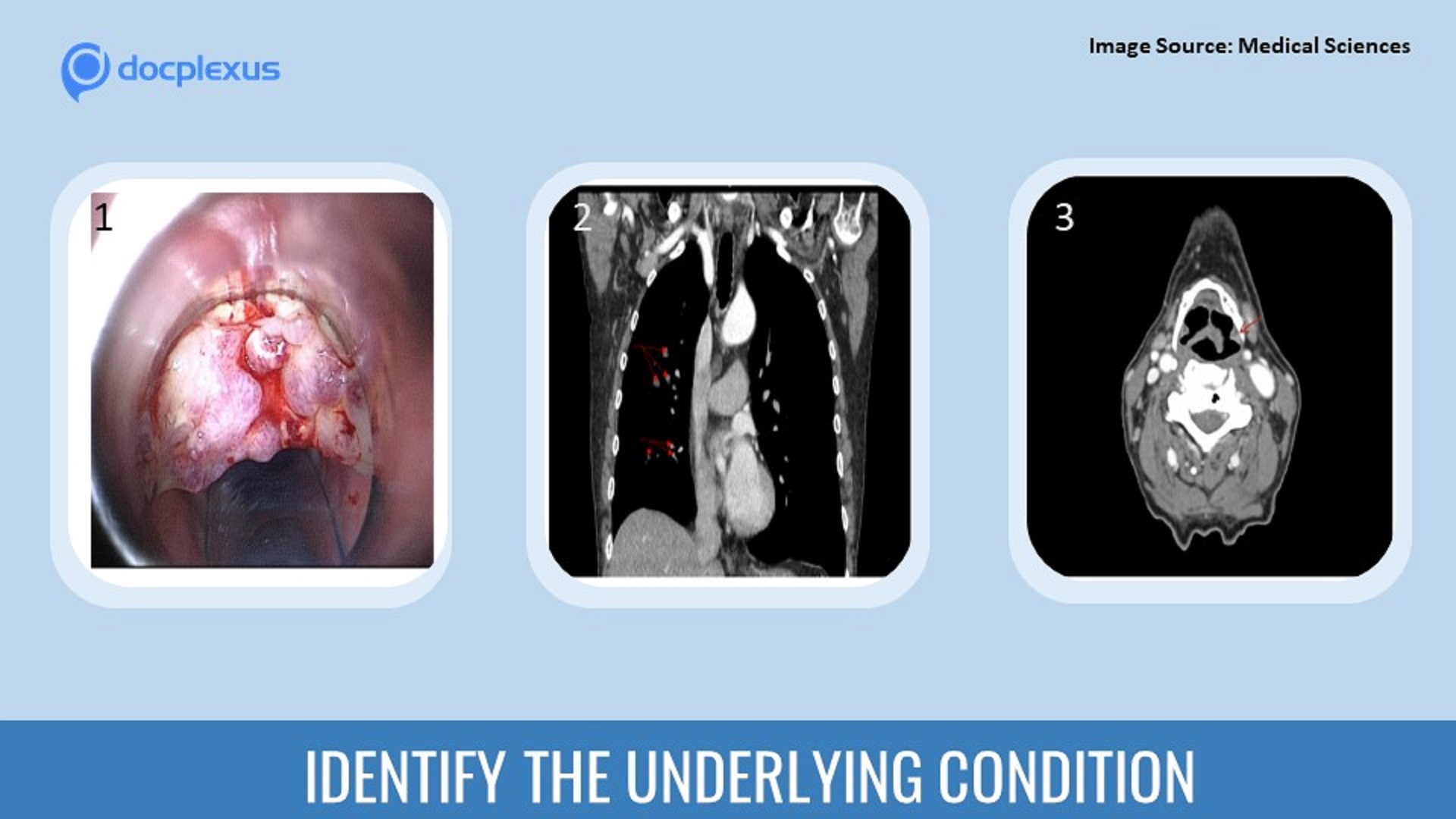
Case presentation A 62-year-old male with a history of a fungal laryngeal infection complained of difficulty in swallowing and weight loss over three months. The patient was a chronic smoker and had an occupational history of being exposed to a dusty environment.
Otorhinolaryngological examination Necrotic masses observed on the laryngeal surface of the epiglottis (Figure 1-Cover image) Vocal folds: mobile and intact Other investigations Chest x-ray: Multiple small foci in the middle lobe and basal part of the right lung Contrast-enhanced CT: Small clustered foci forming a tree-in-bud sign (Figure 2-Cover image) Right lung L1 and S6 has 0.8-1.1 cm size focus Neck-Larynx CT with i/v contrast (Figure 3-cover image): Epiglottis flap appeared thickened Uneven inferior epiglottis contour Growing mass observed Fibrobronchoscopy: Bronchial mucosa was slightly hyperemic. Bronchial sputum was…