
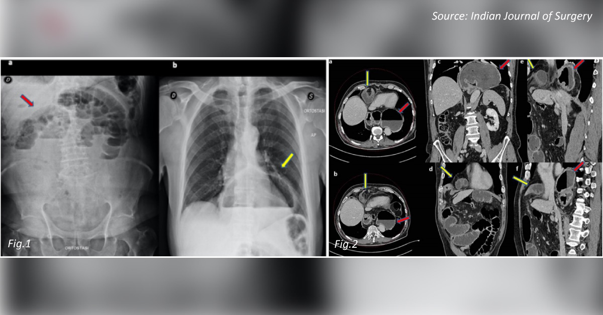
The case An 80-year-old male presented to the emergency department with acute abdominal pain, abdominal distension, nausea, and vomiting. Medical history No significant history was noted. Investigations A classical pattern of mechanical small bowel obstruction was observed after radiographic investigation (Fig 1a). A chest X-ray revealed an air-fluid level in the cardiac shadow (Fig 1b).
A CT scan revealed an extrusion of a small bowel loop in the right hemithorax (Fig 2). Laparoscopy was immediately performed, in which pneumoperitoneum was induced with a veress needle inserted at palmer’s point. Five trocars were used, T1 1 cm above the umbilical scar, T2 in the upper left quadrant, T3 in the upper right quadrant on the midclavicular lines, T4 on the right midclavicular line that is about 10 cm below T2, and T5 below the xiphoid process for liver retraction. * This case is from the…





