
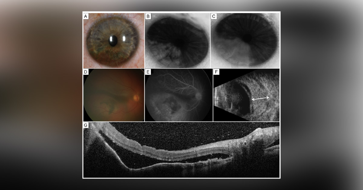
A 2.5-year-old boy presented with amblyopia in the right eye. On examination, he was found to have a pigmented iris “nevus” in the right eye. Visual acuity was found to be counting fingers in the right eye and 20/30 in the left eye. There was no cutaneous or systemic abnormality and no history of Sturge-Weber syndrome . The right eye showed an ill-defined, darkly pigmented iris stromal mass producing corectopia and involving the inferior sector from pupillary margin to the angle, measuring 11 mm in the base and 2 mm in thickness (Image).
The tumor had a rippled surface with thin, atrophic iris stroma draped over large, interweaving intrinsic vessels. Ultrasound biomicroscopy showed a thickened iris but no ciliary body mass. Fluorescein angiography disclosed rapid filling of the mass and no leakage. Fundus examination in the right eye disclosed an ill-defined, orange-colored choroidal…





