
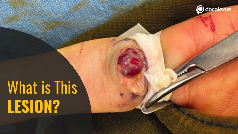
A 53-year-old female presented to the clinic with an asymptomatic, small, protrusive mass on the volar surface of the left index finger at the first phalanx. She reported having first noticed the lesion three months ago. Medical history No significant medical or trauma history Physical examination The lesion had a subcutaneous cystic nodule; no discoloration of the overlying skin It was non-tender and movable. Diagnostic tests Ultrasonography (pre-operative) findings : Cystic lesion of 0.7 × 0.3 × 1.2 cm in size, no vascular increment; suggestive of a ganglionic cyst as shown in figure 1.
Figure 1 . USG showing cystic lesion with no remarkable increased vascularity. Intraoperative findings (Cover image): Location – subcutaneous plane Easily dissectable from the surrounding tissues Soft, red-purple, well-circumscribed cystic mass that looked like hemangioma No remarkable feeding…



