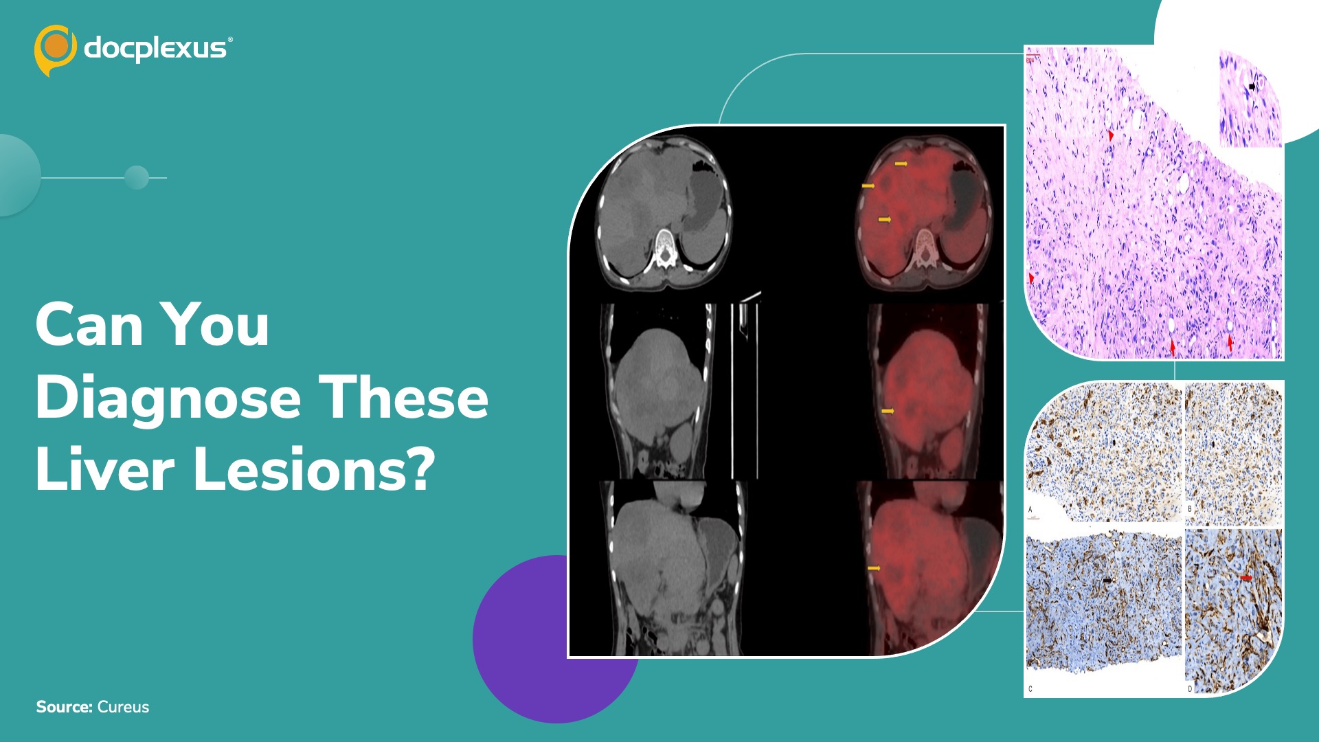
Case presentation A 42-year-old woman was evaluated for persistent generalized weakness following a recent episode of fever and gastrointestinal bleeding, for which she had been managed symptomatically. While the initial symptoms improved, the weakness continued.
She did not report abdominal pain or jaundice. Ultrasonography performed at another hospital Multiple hypoechoic lesions in both hepatic lobes Serum tumor markers Alpha-fetoprotein (AFP), CA 19.9, CEA, and CA-125: Within normal limits Histopathological examination of biopsy sample CT-guided biopsy performed Findings raised the possibility of a malignant epithelial neoplasm Liver tissue core infiltrated by epithelioid cells arranged in cords and vague tubular structures IHC evaluation CK7: Positive in tumor cells CK20, CDX-2, GCDFP-15, GATA3, TTF-1, Napsin-A, WT-1, and PAX8: Negative CK19 and Glypican-3: Negative PET-CT of the…