
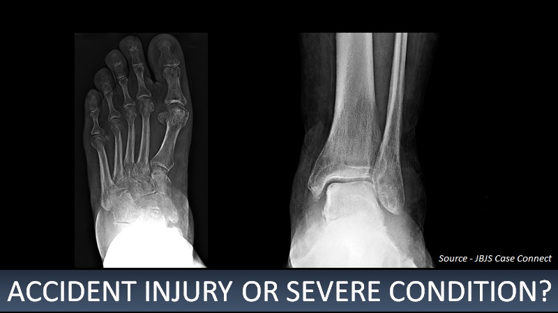
A 58-year-old man presented to the emergency department with a protrusion in the left hindfoot after a fall from standing. Medical history Hypertension Diabetes mellitus complicated by neuropathy Obesity with a body mass index of 42 kg/m2 He had survived a motor-vehicle collision eight months ago, which had led to pain, edema, and numbness in the left hindfoot. Although he was referred to a podiatrist, nothing significant was noted then.
He underwent casting for six weeks, followed by physical therapy. Investigations Magnetic resonance imaging (MRI) revealed the following – Diffuse bone marrow edema in the talus and navicular bones An erosive irregularity at the talonavicular joint A large, multiloculated fluid collection A nuclear scan showed a focal area of uptake in the anterior aspect of the talus and talar head. It also revealed subchondral cysts and moderate to severe…





