
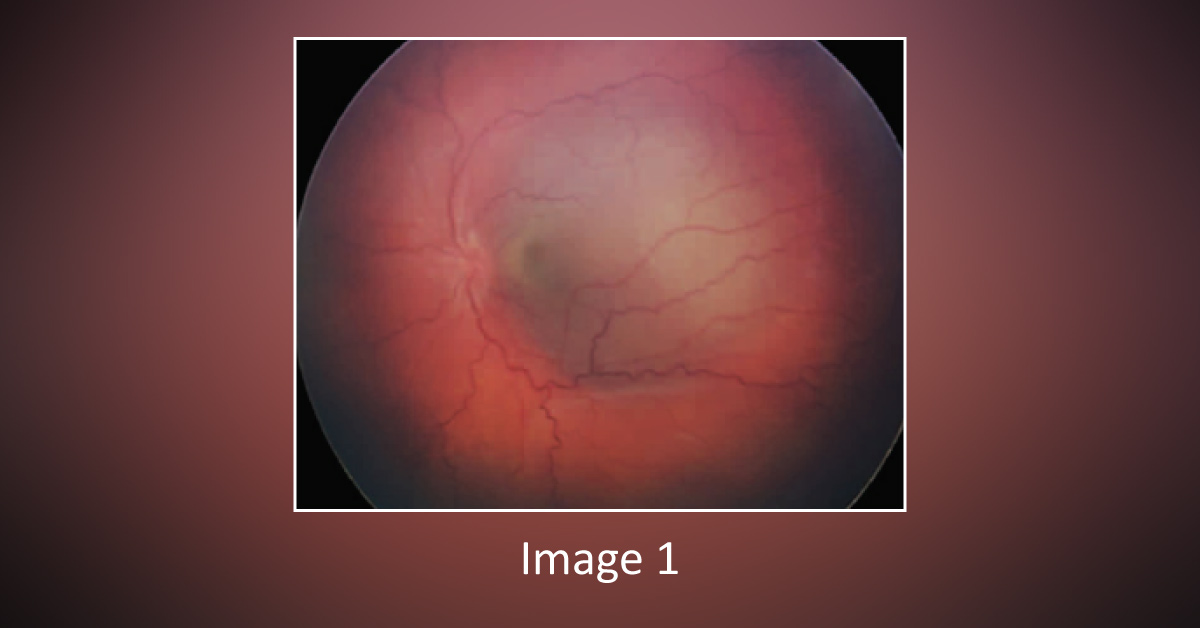
Case presentation A 2-year-old boy was presented for the evaluation of a choroidal mass. The patient had mild periorbital edema and low-grade pain in the left eye for two weeks. Medical history The patient had flu-like symptoms for the last 1.5 weeks, for which he was being treated with ibuprofen with minimal response. Family history Non-contributory Examinations The left eye had mild conjunctival chemosis with periorbital edema and trace eyelid edema, and mild leukocoria.
Dilated ophthalmoscopic examination: A solid gray mass in the macular region, deep into the retina with surrounding subretinal fluid (Image 1). Ultrasonography: The solid mass of 12 mm diameter and 5.1 mm thickness with echodensity (Image 2). Fluorescein angiography: Slight hyperfluorescence of the disc and faint hyperfluorescence of the mass with overlying scattered pinpoint hyperfluorescent leakage. Optical…





