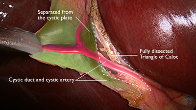
The region known as Calot’s triangle differs today when compared to the area described by Calot in 1890 while he was a medical student. He described in his thesis a triangle bordered by the cystic artery, the cystic duct, and the common hepatic duct. The area described today as his triangle is the region bounded by the cystic duct, common (or right) hepatic duct, and inferior border of the liver.
The change is thought to have occurred because of the practical use of the larger triangle that helps to frame and identify the cystic artery that lies within it. Recognition of critical structures and dissection within Calot’s triangle is of great importance during cholecys- tectomy, especially at the apex of the triangle. The apex of the triangle contains the cystic artery, as discussed, as well as the right branch of the hepatic artery, 95% of accessory right hepatic arteries, and 90% of…