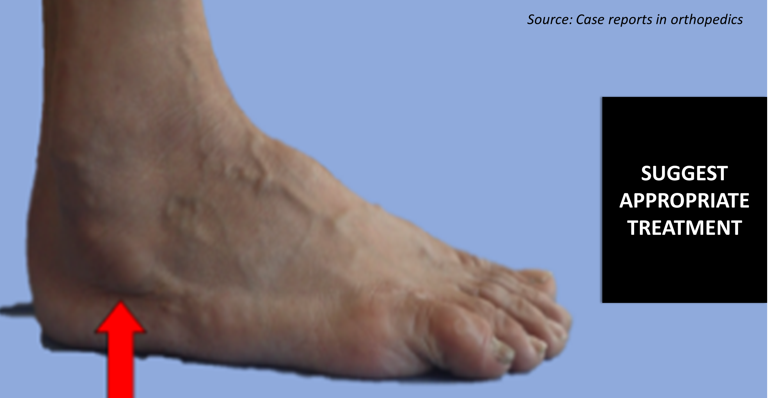
A 63-year-old male patient presented with pain in the lateral aspect of his right hindfoot for the past six years, while walking. Physical examination A palpable bony protrusion on the lateral aspect of the right hindfoot, which was tender (Cover Image, red arrow) Radiographic investigations A routine radiograph and CT scan revealed A bony overgrowth surrounding the peroneal tendon, suggestive of hypertrophic peroneal tubercle (HPT) (Figure 1, red arrow) An ossicle on the outer anterior side of the calcaneus, indicative of the os peroneum (OP) (Figure 1B, yellow arrow) An MRI showed A low intensity lesion on the T1-weighted image around the HPT (Figure 2A, red arrow) A high-intensity lesion on a short T1-inversion recovery (STIR) image surrounding the HPT (Figure 2B, red arrow) Dynamic ultrasonography confirmed HPT and OP causing impacts with active eversion of the foot Pain was…
What Would be Your Recommended Treatment?

Case Challenge
Medical Writer